Review
. 2010 Apr;59(4):547-57.
doi: 10.1136/gut.2009.195131.

Classification and management of refractory coeliac disease

Affiliations
Review

Classification and management of refractory coeliac disease

Alberto Rubio-Tapia et al. Gut. 2010 Apr.

Abstract

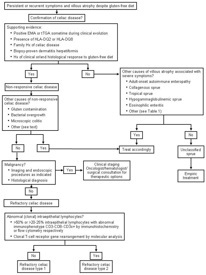
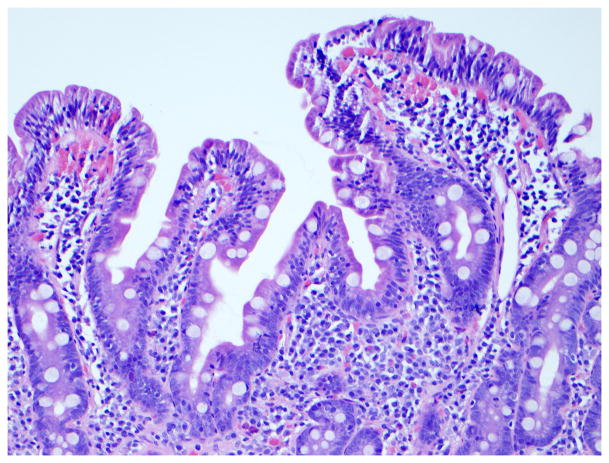
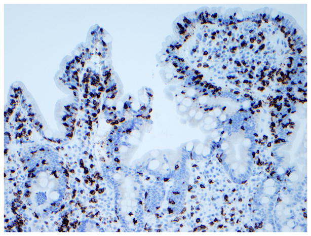
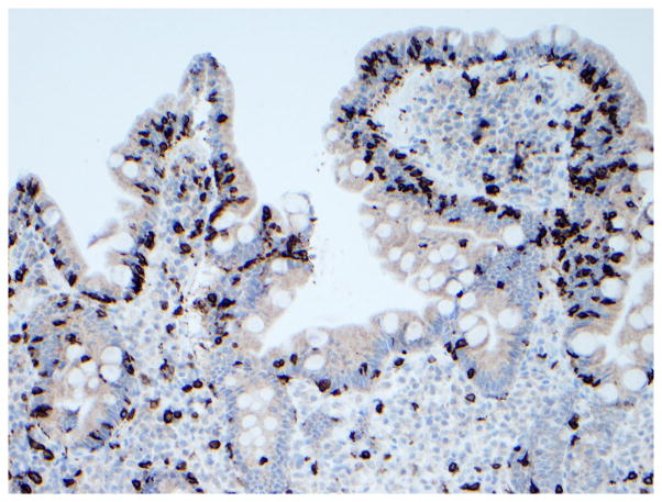
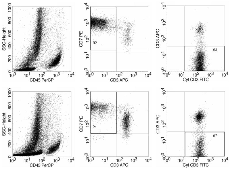
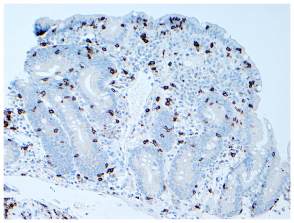
Refractory coeliac disease (RCD) is defined by persistent or recurrent malabsorptive symptoms and villous atrophy despite strict adherence to a gluten-free diet (GFD) for at least 6-12 months in the absence of other causes of non-responsive treated coeliac disease and overt malignancy. Symptoms are often severe and require additional therapeutic intervention besides a GFD. RCD can be classified as type 1 (normal intraepithelial lymphocyte phenotype), or type 2 (defined by the presence of abnormal (clonal) intraepithelial lymphocyte phenotype). Patients with RCD may never have responded to a GFD or may have relapsed despite adherence and initial response to the GFD. RCD type 1 usually improves after treatment with a combination of aggressive nutritional support, adherence to a GFD, and alternative pharmacological therapies. By contrast, clinical response to alternative therapies in RCD type 2 is less certain and the prognosis is poor. Severe complications such as ulcerative jejunitis and enteropathy-associated T cell lymphoma may occur in a subgroup of patients with RCD. The aims of this article are to (1) review recent advances in the diagnosis and management of patients with RCD, and (2) describe current and novel methods for classification of patients with RCD into categories that are useful to predict outcome and direct treatment.


Conflict of interest statement

Competing Interest: Dr. Murray is consultant for ActogeniX Inc., Flamentera Inc., Ironwood pharmaceuticals, and Alvine Inc. Dr Murray is also investigator for ALBA Therapeutics. Dr. Rubio-Tapia declares no competing interest.

Figures

Figure 1
Figure 1. Endoscopic abnormalities in refractory celiac disease
Note the presence of classic endoscopic signs of villous atrophy such as loss of Kerckring’s folds in the duodenum (Panel A), scalloping of circular folds - arrows (Panel B), and fissuring with a mosaic pattern (Panel C). These findings are not specific for refractory celiac disease but excellent predictors of mucosal disease. (Courtesy of Dr. Louis M. Wong Kee Song, Gastroenterology and Hepatology, Mayo Clinic, United States)
Figure 2
Figure 2
Diagnostic Approach in Refractory Celiac Disease
Figure 3
Figure 3. Immunophenotype of intraepithelial lymphocytes in RCD by CD3 and CD8 immunostaining
Panel A, duodenal biopsy specimen from a patient with RCD type 1 (Panel A1, hematoxylin and eosin, 20x original magnification) showing partial villous atrophy and an increased number of intraepithelial lymphocytes with normal immunophenotype characterized by expression of CD3 (Panel A2) and CD8 (Panel A3). Panel B, duodenal biopsy specimen from a patient with type 2 RCD (Panel B1, hematoxylin and eosin, 20x original magnification) showing villous atrophy and abnormal intraepithelial lymphocytes characterized by expression of CD3 (Panel B2) but mostly CD8− (Panel B3). Brown color denotes positive immunostaining (20x original magnification). (Courtesy of Dr. Tsung-Teh Wu, Anatomic Pathology, Mayo Clinic, United States)
Figure 4
Figure 4. Example of flow cytometry analysis of intestinal lymphocytes isolated from duodenal biopsy specimens in a patient with RCD type 2
Upper half: abnormal intraepithelial lymphocytes (92–93%). Botton half: abnormal lamina propria lymphocytes (57%). Left: lymphocyte selection gate based on CD45 positivity and low side scatter. Middle: the abnormal T-cell population by double staining of surface CD3 and CD7 within the CD45+ cells. Right: the abnormal surface CD3−, cytoplasmic CD3+ cells by double staining shown within the CD7+ CD45+ cells. (Courtesy of Dr. Wieke HM Verbeek, VU University Medical Center, Amsterdam, Netherlands)
Figure 5
Figure 5. Histological features of enteropathy-type T-cell lymphoma
Panel A, Low-power magnification of surgical specimen from the jejunum showing subtotal villous atrophy and diffuse infiltration by enteropathy-type T-cell lymphoma in a patient with long-lasting RCD type 2 (hematoxylin and eosin, 4x original magnification). Panel B, Detail of a monotonous tumor cell population with rounded vesicular nuclei, single nucleolus, and abundant cytoplasm (hematoxylin and eosin, 40x original magnification). (Courtesy of Dr. Tsung-Teh Wu, Anatomic Pathology, Mayo Clinic, United States)
Figure 6
Figure 6. Histologic findings in ulcerative jejunitis
Extensive ulceration (left) and villous atrophy (upper right) in the jejunum of a patient with refractory CD compatible with ulcerative jejunitis (Figure 6a, hematoxylin and eosin, 10x original magnification), most intraepithelial lymphocytes in the atrophic mucosa adjacent to ulceration express CD3 (Figure 6b) but not CD8 (Figure 6c). Brown color denotes positive immunostaining (20x original magnification). (Courtesy of Dr. Tsung-Teh Wu, Anatomic Pathology, Mayo Clinic, United States)

References

    1. Di Sabatino A, Corazza GR. Coeliac disease. Lancet. 2009;373:1480–93. - PubMed
    1. Catassi C. The world map of celiac disease. Acta Gastroenterol Latinoam. 2005;35:37–55. - PubMed
    1. Rubio-Tapia A, Kyle RA, Kaplan EL, Johnson DR, Page W, Erdtmann F, Brantner TL, Kim WR, Phelps TK, Lahr BD, Zinsmeister AR, Melton LJ, 3rd, Murray JA. Increased prevalence and mortality in undiagnosed celiac disease. Gastroenterology. 2009;137:88–93. - PMC - PubMed
    1. Lohi S, Mustalahti K, Kaukinen K, Laurila K, Collin P, Rissanen H, Lohi O, Bravi E, Gasparin M, Reunanen A, Maki M. Increasing prevalence of coeliac disease over time. Aliment Pharmacol Ther. 2007;26:1217–25. - PubMed
    1. Green PH, Cellier C. Celiac disease. N Engl J Med. 2007;357:1731–43. - PubMed

Publication types