. 2010 Jun;18(6):1173-82.
doi: 10.1038/mt.2010.13. Epub 2010 Mar 23.

Toward gene therapy for cystic fibrosis using a lentivirus pseudotyped with Sendai virus envelopes

Affiliations

Toward gene therapy for cystic fibrosis using a lentivirus pseudotyped with Sendai virus envelopes

Katsuyuki Mitomo et al. Mol Ther. 2010 Jun.

Abstract

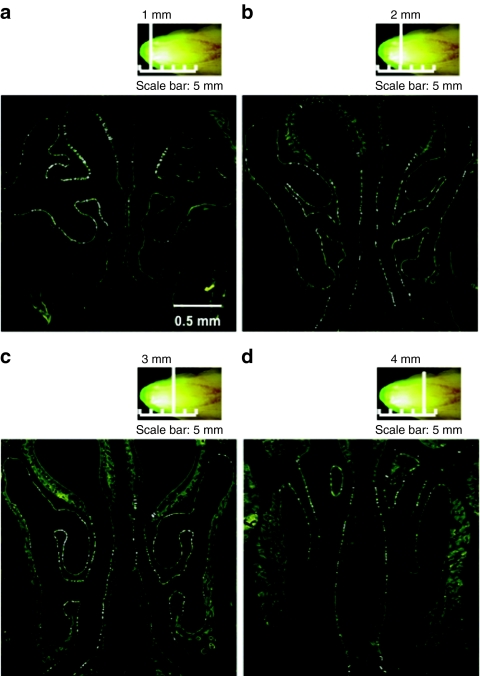
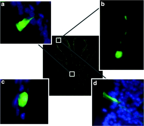
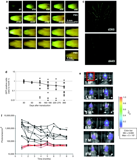
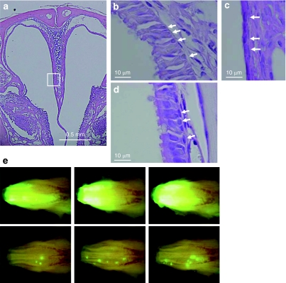
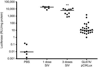
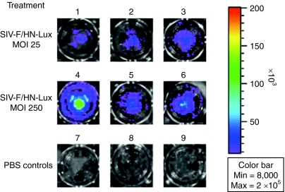
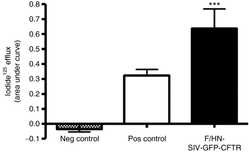
Gene therapy for cystic fibrosis (CF) is making encouraging progress into clinical trials. However, further improvements in transduction efficiency are desired. To develop a novel gene transfer vector that is improved and truly effective for CF gene therapy, a simian immunodeficiency virus (SIV) was pseudotyped with envelope proteins from Sendai virus (SeV), which is known to efficiently transduce unconditioned airway epithelial cells from the apical side. This novel vector was evaluated in mice in vivo and in vitro directed toward CF gene therapy. Here, we show that (i) we can produce relevant titers of an SIV vector pseudotyped with SeV envelope proteins for in vivo use, (ii) this vector can transduce the respiratory epithelium of the murine nose in vivo at levels that may be relevant for clinical benefit in CF, (iii) this can be achieved in a single formulation, and without the need for preconditioning, (iv) expression can last for 15 months, (v) readministration is feasible, (vi) the vector can transduce human air-liquid interface (ALI) cultures, and (vii) functional CF transmembrane conductance regulator (CFTR) chloride channels can be generated in vitro. Our data suggest that this lentiviral vector may provide a step change in airway transduction efficiency relevant to a clinical programme of gene therapy for CF.


Figures

<b>Figure 1</b>
Figure 1
Transduction of mouse nasal epithelium with F/HN-SIV-GFP. The murine nose was perfused in vivo with F/HN-SIV-GFP (4 × 10⁸ TU/mouse) vector and gene expression analyzed 30 days after transduction (n = 8). (a,b) In situ imaging of GFP expression in the nasal cavity. (c,d) Microscopic imaging of GFP in histological sections. The sections were collected (a) 1 mm, (b) 2 mm, (c) 3 mm, and (d) 4 mm into the nasal tissue (vertical white lines). GFP-positive cells appear as small white punctuate signals.
<b>Figure 2</b>
Figure 2
Determination of cell types transduced. The transduced GFP-positive cells were identified using fluorescent microscopy (original magnification ×63) 30 days after administration of F/HN-SIV-GFP (4 × 10⁸ TU/mouse) vector to the mouse nose. (a) Ciliated respiratory epithelial cell, (b) neuronal cell in olfactory epithelium, (c) squamous epithelial cell, (d) non-neuronal cell in olfactory epithelium. The central image shows a cross-section through the mouse nose and white boxes indicate regions in mouse nasal epithelium where respective transduced cell types were found. Panels a, b, and d were rotated ~45°, 130°, and 180° counter clockwise, respectively, to improve clarity of the figure.
<b>Figure 3</b>
Figure 3
Duration of green fluorescent protein (GFP) expression after transduction with F/HN-SIV-GFP. Mouse nasal tissue was perfused with F/HN-SIV-GFP (4 × 10⁸ TU/mouse or phosphate-buffered saline (PBS) and gene expression was analyzed at indicated time points after transduction. (a) Representative in situ images of GFP expression in the nasal cavity from mice analyzed 3–449 days after transduction. (b) In situ imaging of GFP expression in the nasal cavity from 10 mice analyzed 360 days after transduction. (c) Representative microscopic images of GFP expression in histological sections 360 and 449 days after transduction. GFP-positive cells appear as small white punctuate signals. (d) Quantification of transduced cells. GFP-positive cells were quantified on histological sections taken 2 mm into the nasal tissue of the nose. Data from 30 to 360 days after transduction are represented both by mean ± SEM and individual values (ratio to GFP cells positive on day 30). The number (n) per group are 13 (day 30), 3 (day 50), 12 (day 90), 14 (day 160–180), 10 (day 220–270), and 17 (day 360). (e) Bioluminescence in vivo imaging 1 (1 M) to 8 months (8 M) after transduction with F/HN-SIV-lux. Representative images of 2 out of 6 mice are shown. Red box indicates area chosen for quantification of photon emission. (f) Quantification of in vivo bioluminescence over time after transduction with F/HN-SIV-lux (black lines) or PBS (red lines). Each line represents photon emission over time in one animal. ***P < 0.005 compared to bioluminescence one month after gene transfer.
<b>Figure 4</b>
Figure 4
Clustering of transduced cells after the polidocanol-mediated stripping of epithelial cells followed by rapid regeneration. Mouse nasal tissue was perfused with 10 µl of 2% (vol/vol) polidocanol (n = 3). (a) Representative low-power view (original magnification ×50) of the nasal cavity 24 hours after perfusion. Respiratory epithelium, marked by a white box was further magnified (original magnification ×200). The respiratory epithelium before the treatment is shown in b. Arrow indicates basal cells. The respiratory epithelium was completely stripped 24 hours after polidocanol perfusion, whereas the basal cell layer was (c) retained and (d) regenerated 7 days after treatment. (e) This treatment was done after transduction with F/HN-SIV vector. Seven days after transduction of nasal epithelial cells with F/HN-SIV-GFP (4 × 10⁸ transduction units/100 µl/mouse), the nasal epithelium was stripped via perfusion with 10 µl of 2% (vol/vol) polidocanol. Polidocanol treatment was repeated again 3 weeks later. Histological sections were analyzed 58 days after vector administration (30 days after the last polidocanol treatment). In situ imaging of GFP expression in the nasal cavity of untreated mice (top panel in e) or mice treated with polidocanol (bottom panel in e). Clusters of GFP-positive cells were seen in the polidocanol-treated mice. GFP, green fluorescent protein.
<b>Figure 5</b>
Figure 5
Repeat administration of F/HN-SIV to nasal epithelium. Mice were transduced with F/HN-SIV-lux (1 dose) or two doses of F/HN-SIV-GFP (day 0 and day 28) followed by F/HN-SIV-lux 4 weeks later (day 56 = 3 doses). Luciferase expression was measured 30 days after F/HN-SIV-lux transduction and compared to levels achieved with the nonviral gene transfer agent GL67A complexed to a luciferase reporter gene plasmid (pCIKLux). Each dot represents one mouse. Horizontal bars indicate the median per group (**P < 0.01) compared to mice receiving GL67A/plasmid DNA. PBS, phosphate-buffered saline; RLU, relative light units.
<b>Figure 6</b>
Figure 6
Transduction of human air–liquid interface (ALIs) cultures with F/HN-SIV-lux. ALIs were transduced with F/HN-SIV-lux at an approximate multiplicity of infection of 25 (1–3) and 250 (4–6) or treated with phosphate-buffered saline (PBS) (7–9). 5 days after transduction ALIs were treated with luciferin and bioluminescent imaging performed.
<b>Figure 7</b>
Figure 7
Functional confirmation of CF transmembrane conductance regulator (CFTR) production by F/HN-SIV-GFP-CFTR. HEK293T cells were transduced with F/HN-SIV-GFP-CFTR or a control virus carrying green fluorescent protein (GFP) (F/HN-SIV-GFP) at an multiplicity of infection of 500. The iodide efflux assay was performed 2 days after transduction. Cells transfected with an eukaryotic expression plasmid carrying the CFTR complementary DNA under the control of a cytomegalovirus promoter complexed to Lipofectamine 2000 were used as positive control. Data are presented as mean ± SEM. ***P < 0.001 compared to the control virus, n = 6/group. Neg, negative; pos, positive.
<b>Figure 8</b>
Figure 8
Schematic representation of epithelial cell migration in intact and damaged epithelium. (a) Scattered pattern with regeneration in normal condition. (b) Clustered formation after rapid and forced regeneration. We speculate that under normal physiological (undamaged) conditions, turnover may be comparatively slow and that newly generated epithelial cells may move laterally away from the stem or progenitor cell that they originated from a. In contrast, if rapid regeneration is forced (after tissue damage with polidocanol) stem or progenitor cells have to divide rapidly and newly generated epithelial cells may (transiently?) stay in closer proximity to the cell that they originated from b. This hypothesis may explain, why we observed clusters of GFP-positive cells in damaged, but not in undamaged epithelium.

References

    1. Pilewski JM., and , Frizzell RA. Role of CFTR in airway disease. Physiol Rev. 1999;79 1 Suppl:S215–S255. - PubMed
    1. Griesenbach U, Geddes DM., and , Alton EW. Gene therapy progress and prospects: cystic fibrosis. Gene Ther. 2006;13:1061–1067. - PubMed
    1. Yonemitsu Y, Kitson C, Ferrari S, Farley R, Griesenbach U, Judd D, et al. Efficient gene transfer to airway epithelium using recombinant Sendai virus. Nat Biotechnol. 2000;18:970–973. - PubMed
    1. Ferrari S, Griesenbach U, Shiraki-Iida T, Shu T, Hironaka T, Hou X, et al. A defective nontransmissible recombinant Sendai virus mediates efficient gene transfer to airway epithelium in vivo. Gene Ther. 2004;11:1659–1664. - PubMed
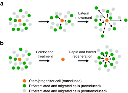
    1. Engelhardt JF. Stem cell niches in the mouse airway. Am J Respir Cell Mol Biol. 2001;24:649–652. - PubMed

Publication types

Substances