. 2010 Jun;18(6):1218-23.
doi: 10.1038/mt.2010.45. Epub 2010 Mar 23.

Dystrophin isoform induction in vivo by antisense-mediated alternative splicing

Affiliations

Dystrophin isoform induction in vivo by antisense-mediated alternative splicing

Sue Fletcher et al. Mol Ther. 2010 Jun.

Abstract

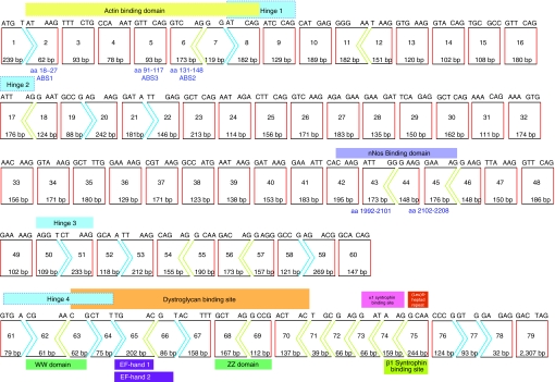
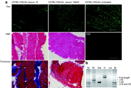
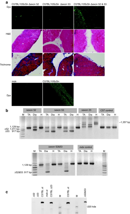
Antisense oligomer-induced manipulation of dystrophin pre-mRNA processing can remove exons carrying mutations, or exclude exons flanking frameshifting mutations, and restore dystrophin expression in dystrophinopathy models and in Duchenne muscular dystrophy (DMD) patients. Splice intervention can also be used to manipulate the normal dystrophin pre-mRNA processing and ablate dystrophin expression in wild-type mice, with signs of pathology being induced in selected muscles within 4 weeks of commencing treatment. The disruption of normal dystrophin pre-mRNA processing to alter the reading frame can be very efficient and offers an alternative mechanism to RNA silencing for gene suppression. In addition, it is possible to remove in-frame exon blocks from the DMD gene transcript and induce specific dystrophin isoforms that retain partial functionality, without having to generate transgenic animal models. Specific exon removal to yield in-frame dystrophin transcripts will facilitate mapping of functional protein domains, based upon exon boundaries, and will be particularly relevant where there is either limited, or conflicting information as to the consequences of in-frame dystrophin exon deletions on the clinical severity and progression of the dystrophinopathy.


Figures

<b>Figure 1</b>
Figure 1
Dystrophin transcript structure,43 showing the reading frame and major functional protein coding domains (actin-binding domain; exons 2–8,20,21,44,45 central rod domain; exons 8–61, which includes nNos binding sites (exons 42–45),46 cysteine-rich domain22 (dystroglycan-binding site; EF1 domain and ZZ domain23) exons 63–70, the α-syntrophin binding site24 and carboxy-terminal domain (exons 70–79). In-frame exons are indicated by rectangles (red sides), whereas codons disrupted by exon junctions are indicated by arrows.
<b>Figure 2</b>
Figure 2
Analysis of dystrophin expression in selected tissues from 10-week-old C57BL/10ScSn mice injected i.p. twice-weekly with peptide-conjugated PMOs (20 mg/kg) targeting dystrophin exons 19 and 20. (a) Immunofluorescent detection of dystrophin on diaphragm cryosections from mice treated with oligomers targeting exon 19 (Δ19) and exons 19 and 20 (Δ19 and 20) (upper panel), haematoxylin and eosin staining (H&E) (middle panel), and Picro Mallory trichrome (lower panel) staining revealing muscle architecture. Sections from sham-treated C57BL/10ScSn and mdx mice are included for comparison (bar = 20 µm). (b) Nested RT-PCR analysis of dystrophin transcripts in tibialis anterior (TA), diaphragm (Dia), and heart (H) from mice treated with oligomers targeting exons 19 and 20 (Δ19 and 20). Transcript product sizes in base pairs (bp) are indicated (M = 100 bp marker). i.p., intraperitoneal; PMO, phosphorodiamidate morpholino oligomer; RT, reverse transcription.
<b>Figure 3</b>
Figure 3
Analysis of dystrophin expression in C57BL/10ScSn mice treated with PMOs (20 mg/kg) targeting dystrophin 52 and 53. (a) C57BL/10ScSn mice were injected twice-weekly with peptide-conjugated PMOs (combined dosage 20 mg/kg) for 4 weeks, beginning at 4 days of age. Dystrophin expression in unfixed cryosections (6 µm) from diaphragm of C57BL/10ScSn mice treated with oligomers targeted to exon 52 (Δ52), exon 53 (Δ53), and exons 52 and 53 together (Δ52 and 53) was detected with Novocastra NCLDYS2 and Zenon Alexafluor 488 (upper panel). Sections from untreated, age-matched C57BL/10ScSn and mdx mice are included for comparison (lowest panel). Sections were also stained with haematoxylin and eosin (H&E) and Picro Mallory trichrome to reveal any pathogenic changes in muscle architecture (middle panels) (bar = 20 µm). (b) Nested RT-PCR across dystrophin exons 49–55 on RNA prepared from diaphragm (Dia), tibialis anterior (TA), and heart (H) of oligomer treated and untreated mice (M = 100 bp marker) and (c) western blot on extracts prepared from diaphragms of C57BL/10ScSn mice treated with oligomers targeted to exon 52 (Δ52), exon 53 (Δ53), and exons 52 and 53 together (Δ52 and 53). Dystrophin was visualized with NCLDYS2 (Novocastra) using Western Breeze (Invitrogen). Samples from diaphragms of C57BL/10ScSn (C57BL ut) and mdx mice (mdx ut) injected with vehicle only (saline), and a normal mouse, treated with an oligomer targeting dystrophin exon 23 (C57BL Δ23) are included for comparison (M = protein standard maker).

References

    1. Wilton SD., and , Fletcher S. Modification of pre-mRNA processing: application to dystrophin expression. Curr Opin Mol Ther. 2006;8:130–135. - PubMed
    1. Deconinck N., and , Dan B. Pathophysiology of duchenne muscular dystrophy: current hypotheses. Pediatr Neurol. 2007;36:1–7. - PubMed
    1. Doran P, Wilton SD, Fletcher S., and , Ohlendieck K. Proteomic profiling of antisense-induced exon skipping reveals reversal of pathobiochemical abnormalities in dystrophic mdx diaphragm. Proteomics. 2009;9:671–685. - PMC - PubMed
    1. Fletcher S, Honeyman K, Fall AM, Harding PL, Johnsen RD, Steinhaus JP, et al. Morpholino oligomer-mediated exon skipping averts the onset of dystrophic pathology in the mdx mouse. Mol Ther. 2007;15:1587–1592. - PubMed
    1. Goyenvalle A, Babbs A, Powell D, Kole R, Fletcher S, Wilton SD, et al. Prevention of dystrophic pathology in severely affected dystrophin/utrophin-deficient mice by morpholino-oligomer-mediated exon-skipping. Mol Ther. 2010;18:198–205. - PMC - PubMed

Publication types

LinkOut - more resources