. 2010 Feb 1:2:140-52.
doi: 10.1093/gbe/evq006.

Mobile regulatory cassettes mediate modular shuffling in T4-type phage genomes

Affiliations

Mobile regulatory cassettes mediate modular shuffling in T4-type phage genomes

Christine Arbiol et al. Genome Biol Evol. .

Abstract

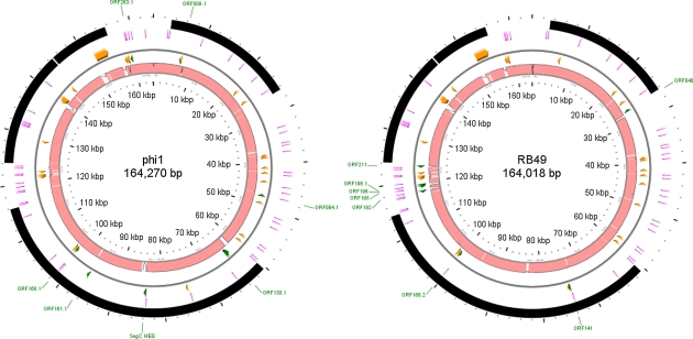
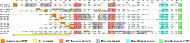
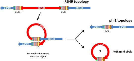
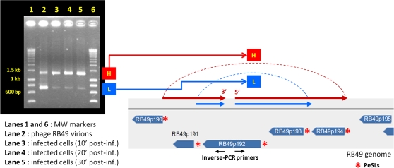
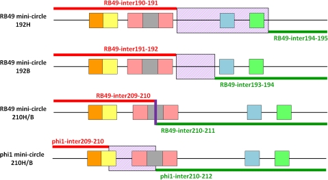
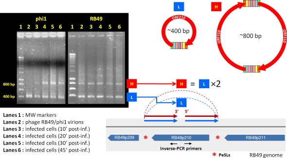
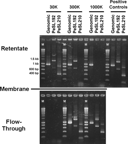
Coliphage phi1, which was isolated for phage therapy in the Republic of Georgia, is closely related to the T-like myovirus RB49. The approximately 275 open reading frames encoded by each phage have an average level of amino acid identity of 95.8%. RB49 lacks 7 phi1 genes while 10 phi1 genes are missing from RB49. Most of these unique genes encode functions without known homologs. Many of the insertion, deletion, and replacement events that distinguish the two phages are in the hyperplastic regions (HPRs) of their genomes. The HPRs are rich in both nonessential genes and small regulatory cassettes (promoter(early) stem-loops [PeSLs]) composed of strong sigma(70)-like promoters and stem-loop structures, which are effective transcription terminators. Modular shuffling mediated by recombination between PeSLs has caused much of the sequence divergence between RB49 and phi1. We show that exchanges between nearby PeSLs can also create small circular DNAs that are apparently encapsidated by the virus. Such PeSL "mini-circles" may be important vectors for horizontal gene transfer.

Keywords: T4-like phage; genome evolution; modular shuffling; regulatory cassette.


Figures

F<sc>IG</sc>. 1.—
FIG. 1.—
Genome comparison of coliphages phi1 and RB49. The inner pink circles represent reciprocal whole-genome alignments of RB49 and phi1 generated by LAGAN (Brudno et al. 2003), the areas indicated in pink shading have a nucleotide homology of >95%. The second and third circles (separated by a thin gray line) are the reverse and forward strands with the divergent (<90% protein identity; orange arrows) and unique (green arrows) genes/ORFs indicated. The fourth circle shows the locations of the PeSLs (radiating magenta lines), and the outermost circle indicates the conserved structural and replication modules (black arcs). The names of the unique genes/ORFs are indicated in green labels outside the circles.
F<sc>IG</sc>. 2.—
FIG. 2.—
A selection of PeSLs from the phi1 genome. The phi1 (and RB49) PeSLs are composed of multiple motifs, starting with the stop codons of the upstream genes (orange) which are typically found in AT-rich regions (pale yellow) located upstream of the -35 and -10 boxes (rose) of the σ70-like promoters. The SLSs (gray stems, with the G-T pairs in bright yellow) are placed in various positions, with the most common positioning being either between the -35 and the -10 boxes or overlapping the -10 box. The PeSLs have at their 3′ extremity a Shine-Dalgarno sequence (blue) and the start codon (green) of the downstream genes. The PeSL sequences were primarily aligned on the basis of the promoter and Shine-Dalgarno sequences.
F<sc>IG</sc>. 3.—
FIG. 3.—
Proposed mechanism for PeSL-mediated modular shuffling. ORF192 in RB49 is flanked by two PeSLs (multicolored rectangles whose constituent elements are color coded as in fig. 2), and a recombination between these two elements, probably in the AT-rich region (marked with an X), has generated a deletion of this ORF from phi1. Such an excision event could also generate a PeSL mini-circle containing ORF192. Note that this process could be reversible and work in the opposite direction to insert ORF192 into phi1 to generate the RB49 topology.
F<sc>IG</sc>. 4.—
FIG. 4.—
PeSL mini-circles created by recombination between neighboring PeSLs in the region of ORF192. Using two primers (the oppositely oriented black arrows) located in the middle of ORF192 in inverse-PCR, two major bands (H: high MW, L: low MW) were detected inside the phage particles and in infected cultures of Escherichia coli after 10 min. Sequencing of these two bands showed that they were circular and contained either two ORFs (192 and 193 for band L) or four ORFs (191–194 for band H), each resulting from an exchange between two pairs of PeSLs (red asterisks) in the vicinity.
F<sc>IG</sc>. 5.—
FIG. 5.—
Schematic diagram of the observed PeSL mini-circles indicating the positions/zones where recombination between the pairs of PeSLs occurred. Alignments of the sequenced PeSL mini-circle sequences compared with the phi1 and RB49 genomes showing the regions of 100% homology on either side of the crossover (red and green lines), as well as the zones (violet hashed box) or exact point (violet line) of exchange events. The different PeSL elements are color coded as in fig. 3.
F<sc>IG</sc>. 6.—
FIG. 6.—
PeSL mini-circles created by recombination between neighboring PeSLs in the region of ORF210. Using two primers (the oppositely oriented black arrows) located in the middle of ORF210 in inverse-PCR, two major bands (H: high MW, L: low MW) could be detected inside phage particles and in infected cultures of Escherichia coli after 10 min. Sequencing of these two bands showed that they were circular and were the products of recombination between the PeSLs flanking ORF210 (red asterisks). The L band contained a monomer of the ORF210 mini-circle sequence, whereas the H band was a dimer of the same sequence.
F<sc>IG</sc>. 7.—
FIG. 7.—
Direct evidence of PeSL mini-circle independent DNA molecules. Extracted RB49 DNA was filtered through three differing pore-size ultrafiltration membranes, which separate larger molecules (remain in retentate) from smaller ones (flow through the filter). The various fractions were used as templates in normal PCR to detect genomic DNA and inverse-PCR to detect the presence of PeSL192 and PeSL210 mini-circles (as in figs. 4 and 6). Lanes labeled with an “M” contain molecular weight markers, with pertinent sizes indicated. The approximate dsDNA cutoffs (defined as 90% retention) for the membranes are as follows: 30K ≈ 150–300 bp; 300K ≈ 1.5–3 kb; 1000K ≈ 5–10 kb.

References

    1. Ackermann HW, Krisch HM. A catalogue of T4-type bacteriophages. Arch Virol. 1997;142:2329–2345. - PubMed
    1. Ausubel FM, et al. Short protocols in molecular biology. New York: J. Wiley and Sons; 1992.
    1. Besemer J, Borodovsky M. GeneMark: web software for gene finding in prokaryotes, eukaryotes and viruses. Nucleic Acids Res. 2005;33:W451–W454. - PMC - PubMed
    1. Botstein D. A theory of modular evolution for bacteriophages. Ann N Y Acad Sci. 1980;354:484–491. - PubMed
    1. Brudno M, et al. LAGAN and MULTI-LAGAN: efficient tools for large-scale multiple alignment of genomic DNA. Genome Res. 2003;13:721–731. - PMC - PubMed