. 2010 Mar 19;6(3):e1000822.
doi: 10.1371/journal.ppat.1000822.

Inhibition of host vacuolar H+-ATPase activity by a Legionella pneumophila effector

Affiliations

Inhibition of host vacuolar H+-ATPase activity by a Legionella pneumophila effector

Li Xu et al. PLoS Pathog. .

Abstract

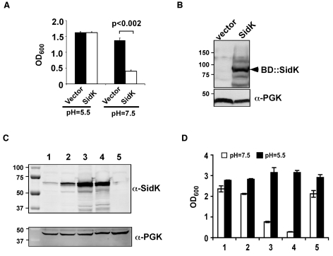
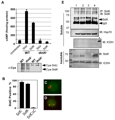
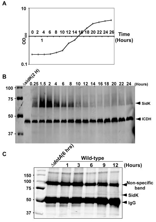
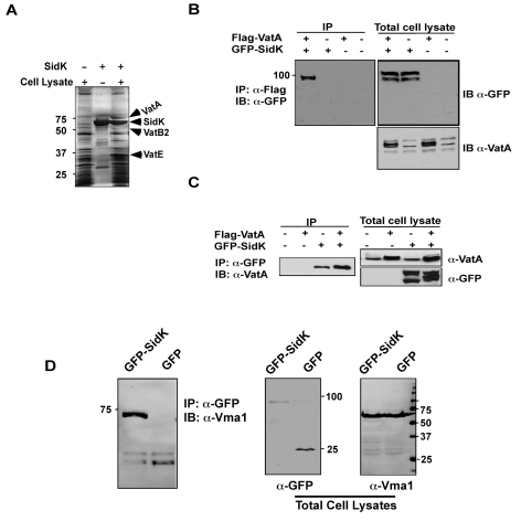
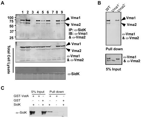
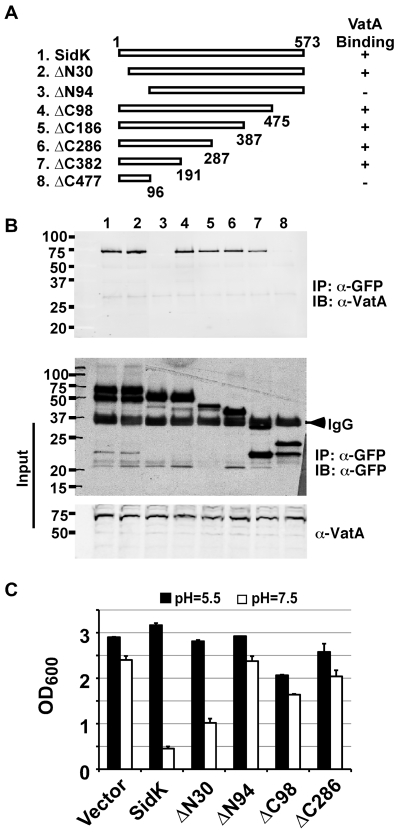
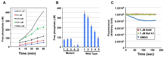
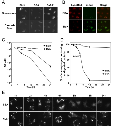
Legionella pneumophila is an intracellular pathogen responsible for Legionnaires' disease. This bacterium uses the Dot/Icm type IV secretion system to inject a large number of bacterial proteins into host cells to facilitate the biogenesis of a phagosome permissive for its intracellular growth. Like many highly adapted intravacuolar pathogens, L. pneumophila is able to maintain a neutral pH in the lumen of its phagosome, particularly in the early phase of infection. However, in all cases, the molecular mechanisms underlying this observation remain unknown. In this report, we describe the identification and characterization of a Legionella protein termed SidK that specifically targets host v-ATPase, the multi-subunit machinery primarily responsible for organelle acidification in eukaryotic cells. Our results indicate that after being injected into infected cells by the Dot/Icm secretion system, SidK interacts with VatA, a key component of the proton pump. Such binding leads to the inhibition of ATP hydrolysis and proton translocation. When delivered into macrophages, SidK inhibits vacuole acidification and impairs the ability of the cells to digest non-pathogenic E. coli. We also show that a domain located in the N-terminal portion of SidK is responsible for its interactions with VatA. Furthermore, expression of sidK is highly induced when bacteria begin to enter new growth cycle, correlating well with the potential temporal requirement of its activity during infection. Our results indicate that direct targeting of v-ATPase by secreted proteins constitutes a virulence strategy for L. pneumophila, a vacuolar pathogen of macrophages and amoebae.


Conflict of interest statement

The authors have declared that no competing interests exist.

Figures

Figure 1
Figure 1. Identification of L. pneumophila protein that inhibits yeast growth in neutral pH medium.
Yeast strains grown to saturation were diluted in medium buffered to the indicated pH at a density of 2×10⁶ cells/ml and the subcultures were incubated at 30°C with vigorous shaking. Cell growth was monitored by measuring OD600 18–24 hrs after establishing the subcultures. A. Growth of a yeast strain expressing SidK fused to the DNA binding domain on pGBKT7. B. Expression of the fusion protein. Yeast strains grown to mid-log phase were lysed with a cracking buffer; SDS-PAGE resolved samples were probed with a SidK specific antibody (upper panel). The 3-phosphoglycerate kinase (PGK) was probed as a loading control (lower panel). Relevant protein size markers (in kDa) are indicated. C. Expression of SidK from different promoters, samples were prepared and probed as described in B (upper panel), the PGK protein was probed as a loading control (lower panel). D. Dose-dependent inhibition of yeast growth in neutral pH medium by SidK. The growth of yeast strains expressing SidK from vectors differing in promoter strength (Materials and Methods) was examined as described in A. Lanes: 1, pADH-SidK(CEN/ARS); 2, pTEF(translation elongation factor 1α)-SidK(CEN/ARS); 3, pTEF-SidK(2 μ); 4, pGPD-SidK(2 μ); 5, pGPD. Data shown are one representative experiment done in triplicates with standard variations shown.
Figure 2
Figure 2. SidK is translocated into infected cells by the Dot/Icm transporter.
A. SidK promoted the translocation of the pertusis toxin Cya into host cells. Differentiated U937 cells were infected with indicated L. pneumophila strains at an MOI of 5 for 1 hr. Cyclic AMP present in lysates of infected cells was measured by ELISA. Lower panel, expression of Cya fusions in L. pneumophila detected with a Cya specific antibody. B–D. Fusion to SidK restores the translocation of the transfer deficient SidΔC100. U937 macrophages were infected with bacteria expressing SidC, SidCΔC100 or SidCΔC100::SidK for one hr; L. pneumophila and SidC was differently labeled by immunostaining. At least 150 vacuoles in triple samples were scored in each experiment. Representative images of vacuoles harboring sidCΔC100 (C) or sidCΔC100::sidK(D); bacteria were labeled in red and SidC was stained in green. E. Dot/Icm-dependent translocation of SidK into infected cells. U937 cells were infected with indicated L. pneumophila strains at an MOI of 5 for 3 hrs. Cleared supernatant obtained with 0.2% saponin was subjected to immunoprecipitation with a SidK specific antibody. Proteins associated with the precipitates were detected by the SidK antibody (upper panel). The Hsp70 in the cell lysates was probed as a loading control and the cytosolic protein ICDH was probed to assess the integrity of the bacterial cells. Note that SidK is present in pellets of all infections except for the deletion mutant (3rd land, lower panel). Lanes: 1, Lp02(dot/icm +); 2, Lp03(dot/icm ); 3, Lp02ΔsidK; 4, Lp02ΔsidK/pSidK. *, bacterial lysate. The sizes (in kDa) of relevant protein markers are labeled on the left side of the blots.
Figure 3
Figure 3. Expression of sidK is induced within hours at the initial phase of bacterial growth.
A. The growth cycle of L. pneumophila in AYE broth. Cultures grown at stationary phase was diluted 1∶20 into fresh medium and the growth of bacteria was monitored by measuring OD600 at indicated time points. B. The expression of SidK at different bacterial growth phase. Lysates were prepared from equal amount of cells withdrawn at the indicated time points and were resolved by SDS-PAGE, the level of SidK was detected by immunoblot with a specific antibody. Lysates of the sidK deletion mutant grown for 2 hrs after dilution was used as a control. The metabolic protein isocitrate dehydrogenase (ICDH) was probed as a loading control. C. Kinetics of SidK translocation during infection. Lysates of U937 cells infected by the dotA mutant or wild type L. pneumophila for indicated time were immunoprecipitated with α-SidK and probed by immunoblott. Note the non-specific band about 100 kDa can serve as a loading control. The sizes (in kDa) of relevant protein markers were labeled on the left side of the blots.
Figure 4
Figure 4. The vacuolar H+-ATPase is the cellular target of SidK.
A. Subunits of the v-ATPase V1 domain were retained by agarose beads coated with SidK. Affigel beads blocked with Tris-HCl buffer (lane 1) or coated with SidK (lane 3) were incubated with mammalian cell lysates. SidK coated beads incubated with lysis buffer (lane 2) served as a second control. After washing with lysis buffer, proteins separated by SDS-PAGE were visualized by silver staining; bands only retained by the SidK coated beads were identified by MALDI/mass spectrometry analysis. B. SidK and VatA form protein complexes in mammalian cells. Lysates of 293T cells transfected to express GFP-SidK or/and Flag-VatA were subjected to immunoprecipitation with an anti-Flag antibody, the presence of SidK in precipitated proteins was detected with GFP specific antibody. C. SidK forms protein complexes with endogenous VatA. Lysates of cells transfected with combinations of plasmids were precipitated with a SidK specific antibody, and proteins bound to beads were detected for VatA. Note that VatA also was precipitated in cells only transfected to express GFP-SidK (lane 3). D. SidK formed complexes with Vma1 of yeast v-ATPase. Lysates of yeast strains expressing GFP or GFP-SidK were coimmunoprecipitated with an anti-GFP antibody, and the presence of Vma1 in the precipitates was detected. In all cases, 5% (50 µg) of total protein was probed as input controls. Relevant protein size markers (in kDa) were indicated.
Figure 5
Figure 5. SidK directly interacts with VatA, the ATP hydrolyzing subunit of v-ATPase.
A. SidK interacts with Vma1 in the absence of other V1 subunits. Lysates of individual vma mutant expressing SidK were coimmunoprecipitated with the anti-SidK antibody and the presence of Vma1 and Vma2 in the precipitates were detected with specific antibodies. Lanes: 1, Δvma4; 2, wild type; 3, Δvma2; 4, Δvma5; 5, Δvma7; 6, Δvma8; 7, Δvma1; 8, Δvma10; 9, Δvma13. The presence of SidK, Vma1 and Vma2 in the samples were probed with 5% proteins used for immunoprecipitation. B. SidK coated beads retained Vma1. Affigel beads coated with SidK were incubated with cell lysates of wild type Δvma1 or Δvma2 mutant. Proteins associated with the beads after extensive wash were probed for Vma1 and Vma2. 5% of lysates was used to probe for the presence of these proteins. Relevant protein size markers (in kDa) were indicated. C. SidK directly interacts with VatA. His6-SidK was incubated with GST-VatA or GST in PBS and potential protein complexes captured with glutathione beads were detected with specific antibody.
Figure 6
Figure 6. SidK binds to VatA via a N-terminal domain.
A. Diagrams of sidK truncation mutants. The numbers at the ends of the bars are the numbers of remaining amino acids for the mutants. +: binds VatA; -, no longer binds VatA. B. Interactions between VatA and the SidK deletion mutants. Lysates of 293T cells expressing each of the mutants fused to GFP were subjected to coimmunoprecipitation with an anti-GFP antibody and the presence of VatA in the precipitates were probed. The middle panel shows protein levels of the mutants probed with a GFP specific antibody after immunoprecipitation. Note that the several mutants that no longer interact with Vma1 still code for stable proteins. Endogenous VatA also was probed as input controls (lower panel). C. Inhibition of yeast growth in neutral pH medium by SidK mutants. Indicated mutants (without any tag) were cloned into p425GPD and yeast growth was assayed as described in Fig. 1.
Figure 7
Figure 7. SidK inhibits v-ATPase-mediated ATP hydrolysis and proton translocation.
A. SidK inhibits ATP hydrolysis in yeast vesicle membranes. BSA, Baf A1 or three different concentrations of His6-SidK was incubated with yeast vesicle membranes, ATP was added to initiate the reaction. The concentration of free phosphate in samples withdrawn at indicated time points was determined by the malachite green method (Materials and Methods). B. SidK specifically inhibits ATPase activity of v-ATPase. Yeast vesicle membranes prepared from wild type or the vma1 mutant were used for similar assays. Note that SidK and the v-ATPase specific inhibitor Baf A1 block ATPase activity in membranes from the wild type but not the vma1 mutant. Lanes (mutant): 1, 1 µM SidK; 2, 1 µM Baf A1; 3, 1 µM BSA; 4, 10 mM EDTA; 5, 1 mM vanadate. Lanes (wild type): 1, 1 µM BSA; 2, 0.1 µM SidK; 3, 0.4 µM SidK; 4, 1 µM SidK; 5, 1 µM Baf A1. C. SidK prevents v-ATPase-mediated proton translocation. ATP was added to reactions containing vacuolar membrane vesicles, acridine orange and the indicated reagents that had been preincubated for 40 min at room temperature. The quenching of acridine orange fluorescence was monitored as described in Experimental Procedures.
Figure 8
Figure 8. Macrophages loaded with SidK are defective in phagosomal acidification and lysosomal digestion of bacteria.
A–B. Phagosomal acidification in macrophages assessed by pH sensitive fluorescein dextran and LysoRed staining. Mouse bone marrow-derived macrophages were loaded with His6-SidK or BSA by syringe loading. Treated cells were incubated with a mixture of fluorescein dextran and cascade blue dextran (0.2 mg/ml) for 1 h, washed 5 times and incubated at 37°C for 4 hrs before being imaged. As a control, Baf A1 (250 nM) was added 40 min before taking the images (A). E. coli cells expressing GFP were incubated with loaded macrophages at an MOI of 20 for 1 hr at 37°C. After incubating with gentamicin for 8 hrs, cells were stained with LysoRed (50 nM) for 15 min. Images were acquired using a fluorescence microscope with identical parameters (B). Bar, 5 µm. C. SidK affects bacterial killing by macrophages. Loaded macrophages were fed with E. coli cells expressing mCherry RFP at an MOI of 10 for 1 hr at 37°C. Samples were treated with gentamicin for 1 h and were extensively washed. Viable E. coli cells were evaluated by plating macrophage lysates on bacteriological media at indicated time points. D. Digestion of bacteria by macrophages. Samples prepared similarly as C were used to evaluate the ratios of macrophages that contain intact E. coli cells. Cells harboring one or more bacterial cells were quantitated at indicated time points. Experiments were performed in triplicates and at least 200 cells were examined each sample. The P values of the relevant data points are indicated. Similar results were obtained in more than three independent experiments. E. Representative images of macrophages fed with fluoresent E. coli cells. Samples at indicated time points were processed and analyzed under a fluorescence microscope and images of typical cells were acquired. Bar, 5 µm.

References

    1. Huynh KK, Grinstein S. Regulation of vacuolar pH and its modulation by some microbial species. Microbiol Mol Biol Rev. 2007;71:452–462. - PMC - PubMed
    1. Ohkuma S, Poole B. Fluorescence probe measurement of the intralysosomal pH in living cells and the perturbation of pH by various agents. Proc Natl Acad Sci U S A. 1978;75:3327–3331. - PMC - PubMed
    1. Shin S, Roy CR. Host cell processes that influence the intracellular survival of Legionella pneumophila. Cell Microbiol. 2008;10:1209–1220. - PubMed
    1. Liu Y, Gao P, Banga S, Luo ZQ. An in vivo gene deletion system for determining temporal requirement of bacterial virulence factors. Proc Natl Acad Sci U S A. 2008;105:9385–9390. - PMC - PubMed
    1. Isberg RR, O'Connor TJ, Heidtman M. The Legionella pneumophila replication vacuole: making a cosy niche inside host cells. Nat Rev Microbiol. 2009;7:13–24. - PMC - PubMed

Publication types

MeSH terms

LinkOut - more resources