. 2010 Mar 19;5(3):e9604.
doi: 10.1371/journal.pone.0009604.

The role of the receptor for advanced glycation end-products in a murine model of silicosis

Affiliations

The role of the receptor for advanced glycation end-products in a murine model of silicosis

Lasse Ramsgaard et al. PLoS One. .

Abstract

Background: The role of the receptor for advanced glycation end-products (RAGE) has been shown to differ in two different mouse models of asbestos and bleomycin induced pulmonary fibrosis. RAGE knockout (KO) mice get worse fibrosis when challenged with asbestos, whereas in the bleomycin model they are largely protected against fibrosis. In the current study the role of RAGE in a mouse model of silica induced pulmonary fibrosis was investigated.

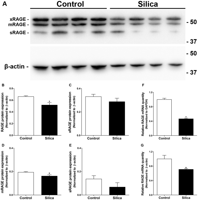
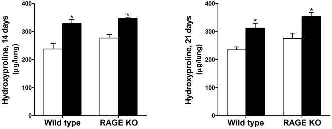
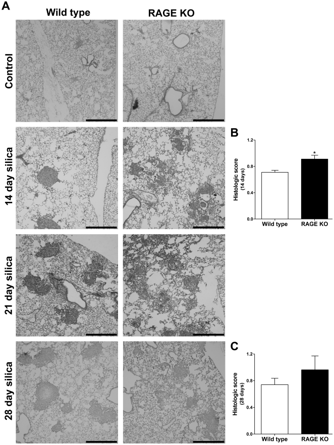
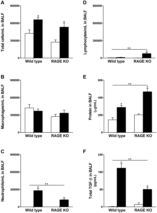
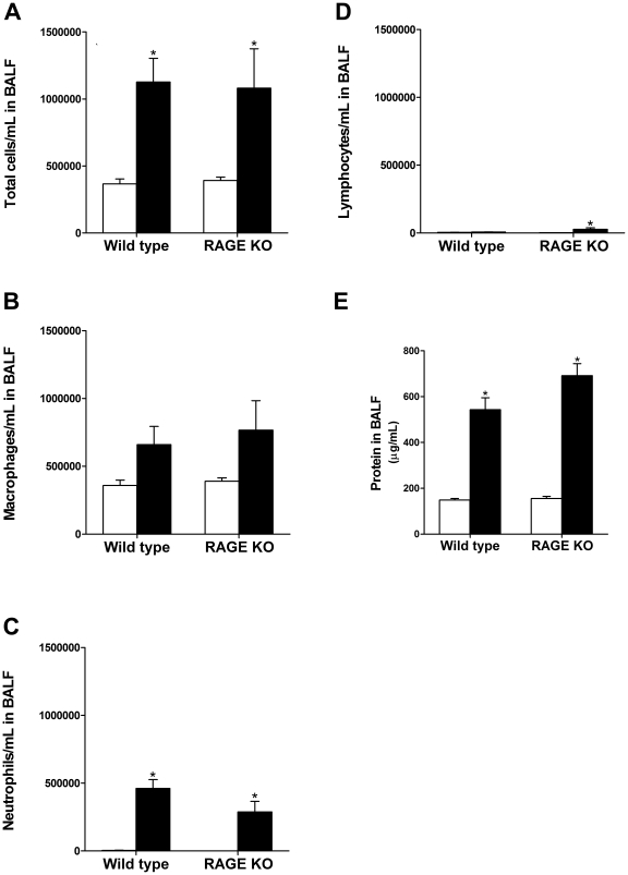
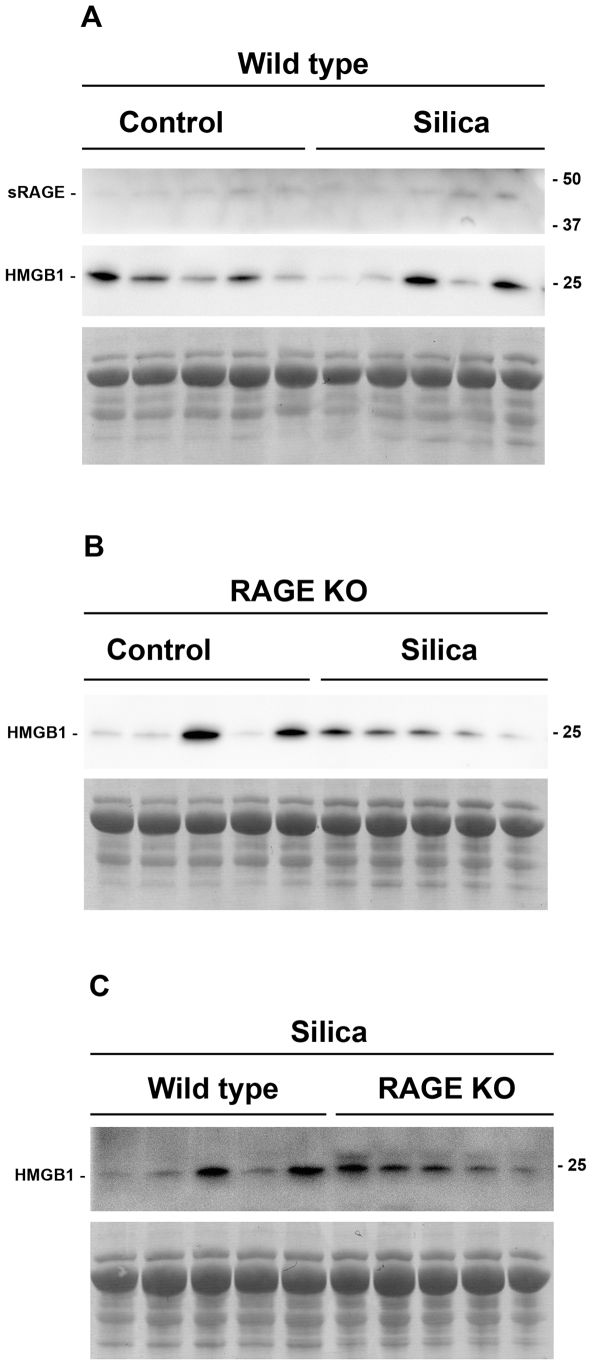
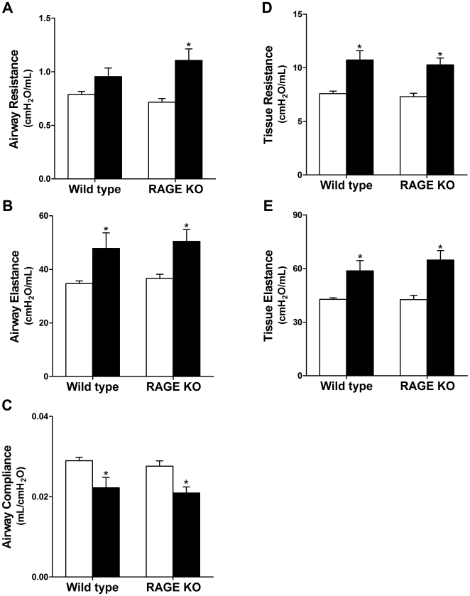
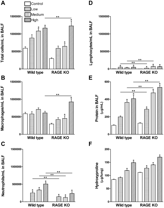
Methodology/principal findings: Wild type (WT) and RAGE KO mice received a single intratracheal (i.t.) instillation of silica in saline or saline alone as vehicle control. Fourteen days after treatment mice were subjected to a lung mechanistic study and the lungs were lavaged and inflammatory cells, protein and TGF-beta levels in lavage fluid determined. Lungs were subsequently either fixed for histology or excised for biochemical assessment of fibrosis and determination of RAGE protein- and mRNA levels. There was no difference in the inflammatory response or degree of fibrosis (hydroxyproline levels) in the lungs between WT and RAGE KO mice after silica injury. However, histologically the fibrotic lesions in the RAGE KO mice had a more diffuse alveolar septal fibrosis compared to the nodular fibrosis in WT mice. Furthermore, RAGE KO mice had a significantly higher histologic score, a measure of affected areas of the lung, compared to WT silica treated mice. A lung mechanistic study revealed a significant decrease in lung function after silica compared to control, but no difference between WT and RAGE KO. While a dose response study showed similar degrees of fibrosis after silica treatment in the two strains, the RAGE KO mice had some differences in the inflammatory response compared to WT mice.

Conclusions/significance: Aside from the difference in the fibrotic pattern, these studies showed no indicators of RAGE having an effect on the severity of pulmonary fibrosis following silica injury.


Conflict of interest statement

Competing Interests: The authors have declared that no competing interests exist.

Figures

Figure 1
Figure 1. RAGE protein and mRNA levels are decreased after silica injury.
(A) Western blot of total protein in lung homogenate of lungs from control and silica treated WT mice, 5 µg of protein were loaded per lane. In addition to mRAGE and sRAGE a third isoform of RAGE, recently termed xRAGE , was also detected by western blot. (B) Protein levels of total RAGE normalized to β-actin revealed a significant decrease in expression after silica injury. (C–E) When split into the different isoforms only mRAGE showed a significant decrease in protein, while both xRAGE and sRAGE had a trend towards a decrease. (F–G) RAGE mRNA levels were significantly decreased after silica injury supporting the observation of the protein levels. This was significant when normalizing to both of the endogenous controls GAPDH and β-actin. Mann Whitney test (n = 4–5 per group). * p<0.05 silica vs. control.
Figure 2
Figure 2. Hydroxyproline levels in WT and RAGE KO lungs were significantly higher in silica treated (closed bars) compared to saline controls (open bars).
There were no significant differences between WT and RAGE KO hydroxyproline levels in the silica injured mice after both 14- and 21 days. Data were analyzed using 2-way ANOVA with a Bonferroni post-test and are means (±SEM) (n = 5–6 per group). * p<0.05 silica vs. control.
Figure 3
Figure 3. RAGE KO mice have a different fibrotic pattern than WT mice.
Lungs were fixed in 10% formalin and paraffin embedded. After H&E staining the lungs were inspected by microscopy (A) and scored according to the degree of fibrosis in each high power field (B and C). When comparing the silica treated WT and RAGE KO lung sections, it is evident that there is a marked difference in the fibrotic pattern among the two strains at both the 14-, 21-, and to a lesser extend the 28 day time point. WT mice have characteristic nodular shaped fibrotic regions, whereas RAGE KO mice develop less dense nodules with a more diffuse alveolar septal fibrosis. Silica treatment resulted in a significant increase in the histologic score for both WT and RAGE KO at the 14- and 28 day time points, and RAGE KO mice had a significantly higher score than WT mice after 14 days (B). After 28 days there was a trend towards what was seen after 14 days (C). Both WT and RAGE KO control treated mice had no fibrosis and therefore a score of 0 throughout (Data not shown). Data are means (±SEM) analyzed by 2-way ANOVA with a Bonferroni post-test. Black scale bars represent 500 µm. (n = 5–6 per group for 14 day time point and n = 3 per group for 28 day time point). * p<0.05 RAGE KO vs. WT.
Figure 4
Figure 4. Analysis of BALF specimens 14 days after treatment.
Lungs were lavaged with 800 µL saline and cells were counted in triplicate. Thirty thousand cells were transferred to a glass slide using a cytospin and stained. Two hundred cells were counted to determine the percentage of macrophages, lymphocytes and neutrophils. (A) Both silica treated WT and RAGE KO (closed bars) had significantly more total cells per mL in the BALF compared to control treated mice (open bars). There was not a significant difference between WT and RAGE KO mice. However, while there was no difference in number of macrophages (B), RAGE KO mice had significantly less neutrophils but more lymphocytes than WT mice treated with silica (C–D). (E) Protein concentration in the BALF was used as a measure of lung permeability and hence lung injury. There was a significant increase in protein concentration in silica treated BALF from both WT and RAGE KO over the controls. In addition, silica treated RAGE KO mice had significantly higher protein concentration in the BALF compared to WT mice. (F) Total TGF-β level in BALF was significantly lower in RAGE KO compared to WT silica treated mice. No active TGF-β was detected in BALF samples. Data are means (±SEM) analyzed by 2-way ANOVA with a Bonferroni post-test. Asterisks above error bars represent comparison to the control treated of the same strain. Asterisks above line represent an interaction and hence a difference between WT and RAGE KO mice. (n = 7–9 per group). * p<0.05 silica vs. control, ** p<0.05 WT vs. RAGE KO.
Figure 5
Figure 5. Analysis of BALF specimens 21 days after treatment.
(A) Both silica treated WT and RAGE KO (closed bars) had significantly more total cells per mL in the BALF compared to control treated mice (open bars). There was not a significant difference between WT and RAGE KO mice. Furthermore, the number of macrophages in both WT and RAGE KO BALF was not significantly increased over control treated mice (B). While the level of neutrophils increased significantly with silica treatment compared to controls for both strains (C), only RAGE KO mice had a significant increase in lymphocytes after silica treatment (D). (E) Protein concentration in the BALF also increased significantly with treatment, but there was no difference between the two strains. Data are means (±SEM) analyzed by 2-way ANOVA with a Bonferroni post-test. (n = 6–7 per group). * p<0.05 silica vs. control.
Figure 6
Figure 6. Western blot of BALF specimens for soluble RAGE and HMGB1.
Equal amounts of protein were loaded in each lane of the individual gels to assure maximum loading of protein. Protein amounts were limited to the maximum amount of protein that could be loaded from the most dilute sample. Protein amounts were as follows; A, 4.4 µg; B, 6.9 µg; C, 11.2 µg. Lower panel in each section shows a section of the ponceau S stained membrane, to assure equal loading of protein. Comparison between blots is not the intension of these western blots. Densitometry analysis was performed on the soluble RAGE and HMGB1 bands, and different treatments compared within each gel by a Mann Whitney test (Data not shown). (A) Compares the two proteins without and with silica treatment in WT mice. Some very weak soluble RAGE bands are visible both in control and silica treated WT BALF. HMGB1 levels were the same in control and silica treated WT mice, and because equal protein was loaded and the fact that the protein concentration in the BALF of WT mice after silica challenge approximately doubled, the overall amount of HMBG1 in the BALF (I.e. amount per volume BALF) is actually increased, this observation was the same for RAGE KO mice (B). (C) Compares HMGB1 levels after treatment with silica in WT and RAGE KO. HMGB1 levels varied to a high degree between samples, and there was no significant difference between the two strains as determined by densitometry analysis.
Figure 7
Figure 7. Lung mechanics after silica injury.
Changes in lung mechanics were similar in WT and RAGE KO mice. Closed bars represent silica treatment and open bars vehicle control. (A) While the airway resistance in silica treated WT mice was not significantly increased over control, the RAGE KO mice treated with silica had a significant increase. All other values (B–E) were significantly changed for both WT and RAGE KO; however there were no other significant differences between WT and RAGE KO. Data are means (±SEM) analyzed by 2-way ANOVA with a Bonferroni post-test. (n = 5–6 per group). * p<0.05 silica vs. control.
Figure 8
Figure 8. Dose response to silica.
(A) The high dose of silica resulted in a significantly higher increase in total cells/mL in the BALF of RAGE KO mice compared to WT mice. (B) RAGE KO mice had more macrophages than WT at the high dose of silica. (C) Neutrophil levels were significantly lower in RAGE KO mice compared to WT mice for all doses of silica. (D) Lymphocyte levels were different among the two strains at the two lower doses of silica. (E) RAGE KO mice had a significant higher level of protein in the BALF after all doses of silica compared to WT mice. (F) Hydroxyproline assay showed no difference in collagen deposition between the two strains. Low; 0.2 mg, medium; 1 mg, high; 5 mg silica. Data are means (±SEM) analyzed by 2-way ANOVA with a Bonferroni post-test. (n = 6–7 per group). * p<0.05 silica vs. control, ** p<0.05 WT vs. RAGE KO.

Similar articles

Cited by

References

    1. Castranova V, Vallyathan V. Silicosis and coal workers' pneumoconiosis. Environ Health Perspect. 2000;108(Suppl 4):675–684. - PMC - PubMed
    1. Kamp DW. Asbestos-induced lung diseases: an update. Transl Res. 2009;153:143–152. - PMC - PubMed
    1. Selman M, King TE, Pardo A. Idiopathic pulmonary fibrosis: prevailing and evolving hypotheses about its pathogenesis and implications for therapy. Ann Intern Med. 2001;134:136–151. - PubMed
    1. Blum RH, Carter SK, Agre K. A clinical review of bleomycin–a new antineoplastic agent. Cancer. 1973;31:903–914. - PubMed
    1. The National Institute for Occupational Safety and Health N. 2008. Silicosis: Number of deaths, crude and age-adjusted death rates, U.S. residents age 15 and over, 1968–2004. Ref. 2007F03-01.

Publication types

Substances