

Longevity of Toxocara cati larvae and pathology in tissues of experimentally infected chickens
- PMID: 20333291
- PMCID: PMC2843852
- DOI: 10.3347/kjp.2010.48.1.79
Longevity of Toxocara cati larvae and pathology in tissues of experimentally infected chickens
Abstract
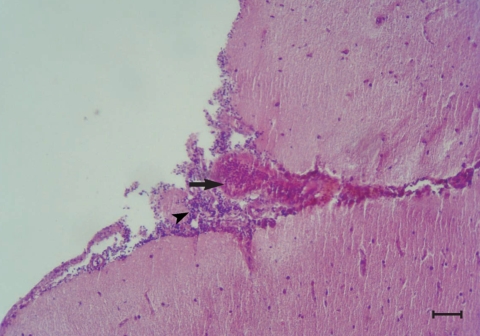
This study was conducted to determine the distribution patterns and duration of stay of Toxocara cati larvae in organs of chickens and to investigate chronic phase and potential zoonotic risk of toxocariasis in chickens. Chickens were orally infected with 1,000 embryonated T. cati eggs and necropsied 240 days post-infection. Organs of the chickens were examined at gross and microscopic levels; tissues were digested to recover larvae. Peribronchiolitis with infiltration of lymphocytes, and hyperplasia of bronchiolar associated lymphatic tissues (BALT) and goblet cells, were evident in the lungs of infected chickens. There were mild hemorrhages and infiltration of lymphocytes and a few eosinophils in the meninges. Larvae were recovered from 30% of the exposed chickens. Larvae recovery indicated that T. cati larvae stay alive for at least 240 days in the chicken brain. Therefore, chickens may potentially act as a paratenic host in nature and transfer T. cati larvae to other hosts.
Keywords: Toxocara cati; chicken; pathology.
Figures

Similar articles
-
Histopathologic changes and larval recovery of Toxocara cati in experimentally infected chickens.Parasitol Res. 2007 Dec;102(1):47-52. doi: 10.1007/s00436-007-0722-5. Epub 2007 Sep 3. Parasitol Res. 2007. PMID: 17768638
-
The effects of benzimidazoles on the larval stage of Toxocara cati in experimentally infected chickens.Trop Biomed. 2009 Apr;26(1):30-9. Trop Biomed. 2009. PMID: 19696725
-
Toxocara cati larvae persist and retain high infectivity in muscles of experimentally infected chickens.Vet Parasitol. 2011 Aug 25;180(3-4):287-91. doi: 10.1016/j.vetpar.2011.03.020. Epub 2011 Mar 21. Vet Parasitol. 2011. PMID: 21482027
-
Toxocara spp. infections in paratenic hosts.Vet Parasitol. 2013 Apr 15;193(4):375-89. doi: 10.1016/j.vetpar.2012.12.033. Epub 2012 Dec 27. Vet Parasitol. 2013. PMID: 23312872 Review.
-
History of Toxocara and the associated larva migrans.Adv Parasitol. 2020;109:17-38. doi: 10.1016/bs.apar.2020.01.037. Epub 2020 Apr 3. Adv Parasitol. 2020. PMID: 32381197 Review.
Cited by
-
Understanding the research and practical needs required to control toxocariasis in Iran.Parasite Epidemiol Control. 2024 Apr 23;25:e00351. doi: 10.1016/j.parepi.2024.e00351. eCollection 2024 May. Parasite Epidemiol Control. 2024. PMID: 38708129 Free PMC article. Review.
-
Nematode larva migrans caused by Toxocara cati in the North Island brown kiwi (Apteryx mantelli).Int J Parasitol Parasites Wildl. 2020 Feb 24;11:221-228. doi: 10.1016/j.ijppaw.2020.02.011. eCollection 2020 Apr. Int J Parasitol Parasites Wildl. 2020. PMID: 32181127 Free PMC article.
-
Detection of larvae of Toxocara cati and T. tanuki from the muscles of free-ranging layer farm chickens.Parasitol Res. 2021 May;120(5):1737-1741. doi: 10.1007/s00436-021-07115-w. Epub 2021 Mar 19. Parasitol Res. 2021. PMID: 33740118
-
Clinical spectrum of symptoms in cerebral Toxocariasis (Review).Exp Ther Med. 2021 May;21(5):521. doi: 10.3892/etm.2021.9953. Epub 2021 Mar 22. Exp Ther Med. 2021. PMID: 33815594 Free PMC article. Review.
-
Natural infection of free-range chickens with the ascarid nematode Toxocara sp.Parasitol Res. 2015 Nov;114(11):4289-93. doi: 10.1007/s00436-015-4669-7. Epub 2015 Aug 29. Parasitol Res. 2015. PMID: 26319520
References
-
- Azizi S, Oryan A, Sadjjadi SM, Zibaei M. Histopathological changes and larval recovery of Toxocara cati in experimentally infected chickens. Parasitol Res. 2007;102:47–52. - PubMed
Publication types
MeSH terms
LinkOut - more resources
Full Text Sources
