. 2010 Mar 19;5(3):e9758.
doi: 10.1371/journal.pone.0009758.

Cleft palate is caused by CNS dysfunction in Gad1 and Viaat knockout mice

Affiliations

Cleft palate is caused by CNS dysfunction in Gad1 and Viaat knockout mice

Won-Jong Oh et al. PLoS One. .

Abstract

Background: Previous studies have shown that disruption of GABA signaling in mice via mutations in the Gad1, Gabrb3 or Viaat genes leads to the development of non-neural developmental defects such as cleft palate. Studies of the Gabrb3 and Gad1 mutant mice have suggested that GABA function could be required either in the central nervous system or in the palate itself for normal palatogenesis.

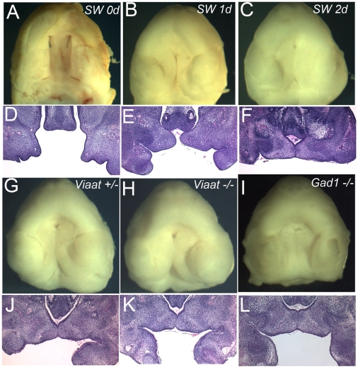
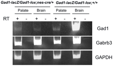
Methodology/principal findings: To further examine the role of GABA signaling in palatogenesis we used three independent experimental approaches to test whether Gad1 or Viaat function is required in the fetal CNS for normal palate development. We used oral explant cultures to demonstrate that the Gad1 and Viaat mutant palates were able to undergo palatogenesis in culture, suggesting that there is no defect in the palate tissue itself in these mice. In a second series of experiments we found that the GABA(A) receptor agonist muscimol could rescue the cleft palate phenotype in Gad1 and Viaat mutant embryos. This suggested that normal multimeric GABA(A) receptors in the CNS were necessary for normal palatogenesis. In addition, we showed that CNS-specific inactivation of Gad1 was sufficient to disrupt palate development.

Conclusions/significance: Our results are consistent with a role for Gad1 and Viaat in the central nervous system for normal development of the palate. We suggest that the alterations in GABA signaling lead to non-neural defects such as cleft palate as a secondary effect due to alterations in or elimination of fetal movements.


Conflict of interest statement

Competing Interests: The authors have declared that no competing interests exist.

Figures

Figure 1
Figure 1. Targeted disruption of the Viaat gene in mice.
(A) A schematic representation of the Viaat wild-type genomic locus (Genomic Structure), the targeting vector (Targeting Construct), and the mutant locus (Targeted structure) are shown. A LacZ sequence was inserted into an NcoI site at the Viaat start codon. The lox sites that flank the Neo resistance cassette are indicated with two arrowheads in the region immediately 3′ to the lacZ sequence. The locations of the 5′ (5′ Probe) and 3′ (3′ Probe) flanking probes used in characterizing the Viaat allele are indicated above the map of the wild type Viaat locus. The positions of the restriction sites used in the Southern blot analysis of genomic DNA from ES cells are indicated by single letters. Restriction sites are indicated as follows: H (HindIII), Nc (NcoI), A (Asp718) and N (NotI). (B) Southern blot analysis of genomic DNA from a wild type parental ES cell line (+/+) and a targeted ES cell line used to generate the ViaatlacZ mouse (+/-) are shown. In genomic DNA digested with Asp718 the 3′flanking probe hybridized to a 15.5 kb wild type fragment and a 10.9 kb from the targeted allele (Asp718 panel) and in genomic DNA digested with HindIII this probe hybridized to a 12.1 kb wild type band and a 9.8 kb mutant band (HindIII panel). (C) RT-PCR analysis of cDNA from wild-type (+/+) , heterozygous (+/−), and mutant embryo brain at E16.5. For each genotype RT-PCR was performed on RNA that had been reverse transcribed (+ lanes) and RNA that had not been incubated with reverse transcriptase (−) to control for non-specific amplification. GAPDH primers were used as a positive control. (D) Localized expression of β-galactosidase activity in ViaatlacZ and Gad1lacZ heterozygotes. The panels from left to right show β-galactosidase activity in E11.5, and E14.5 ViaatlacZ embryos and in E12.5 and E14.5 Gad1lacZ embryos.
Figure 2
Figure 2. Viaat mutants exhibit cleft palate and umbilical hernia.
(A) Gross morphology of a ViaatlacZ (−/−) mutant at E18.5 with hunched posture as compared to a wild type E18.5 littermate (+/+). (B, C) Examination of E17.5 embryos showed that all of the ViaatlacZ mutants exhibited a cleft palate (C) as compared to wild type (B). (D) Viaat mutants have small subcutaneous bumps on the dorsal side of cervical region. (E, F) Compared to wild type (E) nearly all of the ViaatlacZ mice exhibited an umbilical hernia (F). (G) Gad1lacZ mutant E17.5 mice also displayed a similar hernia phenotype.
Figure 3
Figure 3. Viaat mutants exhibit delays in palate shelf elevation.
(A-D) H&E stained coronal sections showing normal palatogenesis in wild type E13.5, E14.5, E15.5 and E18.5 mice. (E-H) Coronal sections of ViaatlacZ homozygous mutants at E13.5, E14.5, E15.5 and E18.5, showing a failure of palate shelf elevation in the mutants. P, palate; PS, palatal shelf; OC, oral cavity; T, tongue.
Figure 4
Figure 4. The developing palatal shelves do not express Viaat transcripts.
RNA from dissected E13.5 and E14.5 brain tissue (Brain) or E13.5 and E14.5 dissected palate shelves (Palate) was analyzed by RT-PCR using primers corresponding to the Viaat, Gad1, Gabrb3 and Gapdh coding sequences. For each tissue and stage RT-PCR was performed with cDNA (+) or with RNA that had not been reverse transcribed (−) to control for non-specific amplification. Gapdh was used as a positive control.
Figure 5
Figure 5. In vitro fetal palate explant culture.
(A-F) Wild-type palates from Swiss Webster (SW) embryos at E13.5 were dissected and cultured for 2 days. (A-C) View of the oral surface of explants prior to culture (A) and after 1 day (B) or 2 days (C) of culture. (D-F) sections through the palate explants in shown in panels A-C. (G-I) Normal palatogenesis of the Viaat heterozygous (+/−) (G) and homozygous (−/−) (H) explants after 2 days in culture. Palate explants from the Gad1 mutant embryo at E13.5 also developed normally during culture for 2 days (I). (J-L) Sections of the palate explants shown in panels G-I.
Figure 6
Figure 6. Specific inactivation of Gad1 in the CNS of Gad1flox/Gad1lacZ NesCre E14.5 day old embryos.
RT-PCR analysis of RNA extracted from dissected palate shelves (Palate) or brain (Brain). The genotype of the tissue is indicated above. For each sample PCR amplification was performed with cDNA (+ lanes) as well as RNA that had not been reverse transcribed (- lanes).

Similar articles

Cited by

References

    1. Culiat CT, Stubbs LJ, Woychik RP, Russell LB, Johnson DK, et al. Deficiency of the beta 3 subunit of the type A gamma-aminobutyric acid receptor causes cleft palate in mice. Nat Genet. 1995;11:344–346. - PubMed
    1. Homanics GE, DeLorey TM, Firestone LL, Quinlan JJ, Handforth A, et al. Mice devoid of gamma-aminobutyrate type A receptor beta3 subunit have epilepsy, cleft palate, and hypersensitive behavior. Proc Natl Acad Sci U S A. 1997;94:4143–4148. - PMC - PubMed
    1. Condie BG, Bain G, Gottlieb DI, Capecchi MR. Cleft palate in mice with a targeted mutation in the gamma-aminobutyric acid-producing enzyme glutamic acid decarboxylase 67. Proc Natl Acad Sci U S A. 1997;94:11451–11455. - PMC - PubMed
    1. Asada H, Kawamura Y, Maruyama K, Kume H, Ding RG, et al. Cleft palate and decreased brain gamma-aminobutyric acid in mice lacking the 67-kDa isoform of glutamic acid decarboxylase. Proc Natl Acad Sci U S A. 1997;94:6496–6499. - PMC - PubMed
    1. Wojcik SM, Katsurabayashi S, Guillemin I, Friauf E, Rosenmund C, et al. A shared vesicular carrier allows synaptic corelease of GABA and glycine. Neuron. 2006;50:575–587. - PubMed

Publication types