. 2010 Mar 19;5(3):e9777.
doi: 10.1371/journal.pone.0009777.

Cyclotraxin-B, the first highly potent and selective TrkB inhibitor, has anxiolytic properties in mice

Affiliations

Cyclotraxin-B, the first highly potent and selective TrkB inhibitor, has anxiolytic properties in mice

Maxime Cazorla et al. PLoS One. .

Abstract

In the last decades, few mechanistically novel therapeutic agents have been developed to treat mental and neurodegenerative disorders. Numerous studies suggest that targeting BDNF and its TrkB receptor could be a promising therapeutic strategy for the treatment of brain disorders. However, the development of potent small ligands for the TrkB receptor has proven to be difficult. By using a peptidomimetic approach, we developed a highly potent and selective TrkB inhibitor, cyclotraxin-B, capable of altering TrkB-dependent molecular and physiological processes such as synaptic plasticity, neuronal differentiation and BDNF-induced neurotoxicity. Cyclotraxin-B allosterically alters the conformation of TrkB, which leads to the inhibition of both BDNF-dependent and -independent (basal) activities. Finally, systemic administration of cyclotraxin-B to mice results in TrkB inhibition in the brain with specific anxiolytic-like behavioral effects and no antidepressant-like activity. This study demonstrates that cyclotraxin-B might not only be a powerful tool to investigate the role of BDNF and TrkB in physiology and pathology, but also represents a lead compound for the development of new therapeutic strategies to treat brain disorders.


Conflict of interest statement

Competing Interests: The authors have declared that no competing interests exist.

Figures

Figure 1
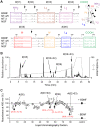
Figure 1. Purification and identification of binding determinants of BDNF.
(A) Alignment of the amino-acid sequences of rat BDNF, NT-4/5, NT-3 and NGF (numbered based on BDNF sequence, dashes represent gaps, points represent identities and stars represent strongly similar amino acids). Variable regions are boxed and labeled (Loops L1, L2, L3 and L4). (inset) Three-dimensional structure of a BDNF monomer (PDB entry, 1bnd). Isolation of these regions by endoproteinase Glu-C V8 (black arrows) resulted in the production of six fragments (#(01) to #(06)). (B) Fragments were purified by HPLC using a non-linear gradient (dashed line) and identified by ESI-MS (solid line). All expected fragments and fragments resulting from miscleavage of BDNF were found. Only the four fragments capable of inhibiting the BDNF-induced TrkB activity in (C) are noted. (C) Representative KIRA-ELISA inhibition profile of 80 HPLC fractions. Fractions (∼0.3 µM final) were assayed in the presence of 1 nM BDNF. Most fractions did not produce significant inhibitions (○) except four fractions (• single fragments, ▴ miscleavage fragments). Mean ± s.e.m. of values obtained in triplicate in 8 independent experiments are noted in brackets.
Figure 2
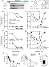
Figure 2. Cyclotraxin-B is a highly potent allosteric inhibitor of TrkB receptor with long-lasting effects.
(A) Design of cyclotraxin-B. (Left) Sequence alignment of the four neurotrophins on the highly variable region III (gray box). The two cysteine residues used for cyclization are in red. Point, conserved residues; asterisk, highly similar residues. (Right) 3-D structure of a BDNF monomer (adapted from PDB entry 1bnd) and sequence of cyclotraxin-B. Variable regions are indicated and higher magnification of region III is boxed. The position of the disulfide bond is shown as a dashed line. (B-E) Characterization of TrkB inhibition by cyclotraxin-B using KIRA-ELISA assays in TetOn-rhTrkB cells (B,C) and in cortical neurons (D,E). (B,D) Increasing concentrations of cyclotraxin-B (B, n = 6; D, n = 5) or L2-8 (n = 3) were added to the cells with or without BDNF. (C,E) BDNF concentration-response experiments with or without cyclotraxin-B (C, 1 µM, n = 6; E, 100 nM, n = 6). Addition of cyclotraxin-B resulted in a significant uncompetitive antagonism (C, F1,285 = 88.0, P<0.0001; E, F1,279 = 199.9, P<0.0001) and did not change BDNF EC50 (C, BDNF 672±92 pM, + cyclotraxin-B 749±91 pM; E, BDNF 186±50 pM, + cyclotraxin-B 178±52 pM), as shown by Eadie-Hofstee plotting of the data (insets). Results are expressed as the ratio between phospho- and total-TrkB in percentage of basal value. Data are mean ± s.e.m. (triplicates, n = 6), except for insets where data are mean. (F) Slow reversibility of cyclotraxin-B inhibition in TetOn-rhTrkB cells and in cultured neurons. After 30-min exposure to cyclotraxin-B, cells were rapidly washed and incubated in KIRA-ELISA medium for increasing times before the addition of BDNF. Data are mean ± s.e.m. (triplicates, n = 4) and are expressed in percentage of inhibition. (G) Means ± s.e.m. of half times obtained in (F). * P = 0.02.
Figure 3
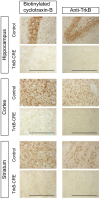
Figure 3. Cyclotraxin-B interacts with TrkB.
Fixed slices from adult control and transgenic CamKIIa-CRE x TrkB flox/flox (TrkB-CRE) mice were incubated overnight with biotinylated cyclotraxin-B or anti-TrkB antibody. Regions of the forebrain in which the expression of TrkB is knocked out in the transgenic mice are shown (Left to right: dentate gyrus of hippocampus, prefrontal cortex, dorsal striatum; 40× magnification). Data presented for biotinylated cyclotraxin-B are those obtained with 0.1 µM of but are similar with concentrations up to 1 µM. Scale bar, 200 µm.
Figure 4
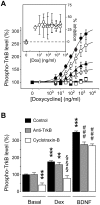
Figure 4. Cyclotraxin-B inhibits both BDNF-dependent and -independent TrkB activity.
(A) Cyclotraxin-B inhibition of density-related TrkB activity. Cells were induced with doxycycline (see Fig. S4B) and were treated with BDNF (4 nM), cyclotraxin-B (1 µM) and K252a (1 µM) before KIRA-ELISA analysis (• BDNF, ○ BDNF+cyclotraxin-B, ▴ basal, Δ cyclotraxin-B, ▪ K252a, □ K252a+cyclotraxin-B). Inset shows amplitude of inhibition of BDNF-induced and basal activities. Data are mean ± s.e.m. (triplicates, n = 3). Results are expressed in percentage of values obtained in non-induced cells. (B) Cyclotraxin-B inhibition of glucocorticoid-dependent TrkB activity in neurons. Cortical neurons were treated with control medium, dexamethasone (1 µM, 2 h) or BDNF (4 nM), in presence or not of cyclotraxin-B (1 µM) or a monoclonal anti-TrkB antibody (30 µg/ml). Data are mean ± s.e.m. (triplicates, n = 6) and are expressed in percentage of basal values; ** P<0.01, *** P<0.001, compared to basal/control condition; §§§ P<0.001, compared to dexamethasone alone; ### P<0.001, compared to BDNF condition.
Figure 5
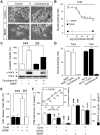
Figure 5. Cyclotraxin-B inhibits normal and deleterious cellular signaling events associated with TrkB but not TrkA nor TrkC.
(A-D) Cyclotraxin-B inhibits TrkB- but not TrkA- nor TrkC-dependent neurite outgrowth. (A) Representative photomicrographs of nnr5 PC12-TrkB cells treated for 48 h with cyclotraxin-B and/or BDNF. 20× magnification. (B) Quantitative analysis of BDNF-induced neurite outgrowth in presence of increasing concentrations of cyclotraxin-B. Data are mean ± s.e.m. (sixplicates; n = 5). (C) Quantitative analysis and representative western blot of total and phospho-MAPK in nnr5 PC12-TrkB cells treated as indicated. Data are mean ± s.e.m. (n = 4) and are expressed in percentage of basal condition; *** P<0.001 compared to control; §§§ P<0.001, compared to BDNF. (D) nnr5 PC12-TrkA and -TrkC cells were incubated with cyclotraxin-B (1 µM) in presence or not of NGF or NT-3, respectively. (E) Cyclotraxin-B prevents BDNF-induced neurons death. Cortical neurons were treated with cyclotraxin-B or BDNF, as described in methods. Data are mean ± s.e.m. (sixplicates, n = 5) expressed in percentage of control. *** P<0.001 compared to control; §§§ P<0.001, compared to BDNF. (F) Cyclotraxin-B inhibits cap-dependent protein translation in cortical neurons. [35S]-methionine incorporation into proteins (white bars) was measured in cortical neurons exposed to cyclotraxin-B, K252a and BDNF, as indicated. Phosphorylation of eIF4E was quantified by immunoblots (black bars). Data are mean ± s.e.m. (triplicates, n = 5) and are expressed in percentage of basal condition; * P<0.05, ** P<0.01, *** P<0.001, compared to respective basal condition; § P<0.05, §§ P<0.01, compared to respective BDNF condition. Inset shows correlations between TrkB-dependent protein synthesis and eIF4E phosphorylation. (1) K252a, (2) cyclotraxin-B, (3) basal, (4) BDNF + cyclotraxin-B, (5) BDNF.
Figure 6
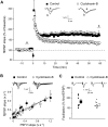
Figure 6. Cyclotraxin-B impairs HFS-induced LTP without affecting basal synaptic transmission.
(A) Effect of prior exposure to cyclotraxin-B on tetanus-induced LTP in the CA1 area. Grouped recordings are shown before and after tetanic stimulation (arrow) for control and cyclotraxin-B-treated hippocampal slices. Representative fEPSPs recorded 5 min before (a) and 60 min after (b) LTP induction are shown. Results are expressed in percentage of baseline response and are the mean ± s.e.m. (n = 8−9 slices). (B) Input-output curves plotting of fEPSP slopes against presynaptic fiber volley (PSFV) slope in control (r 2 = 0.84; n = 7) and cyclotraxin-B-treated slices (r 2 = 0.92; n = 10). Individual traces obtained for control and cyclotraxin-B-treated slices are shown. (C) Scatter plot depicting the facilitation ratio obtained in control (n = 7) and cyclotraxin-B-treated slices (n = 10). Mean ± s.e.m. is indicated by the large symbol with the error bars while smaller symbols represent data obtained in each experiment. Representative sweeps obtained with control and cyclotraxin-B-treated slices are shown.
Figure 7
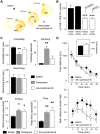
Figure 7. Cyclotraxin-B is active in vivo after systemic injections and demonstrates anxiolytic-like but not antidepressant-like effects.
(A) Brain localization of biotinylated tat-cyclotraxin-B after intravenous injections. Cx, cortex; St, striatum; NAc, Nucleus Accumbens; Hip, hippocampus. (B) KIRA-ELISA analysis of brain TrkB receptors from mice treated with saline (sal), tat-empty (empty) or tat-cyclotraxin-B (cyclo) as described in Figure S8. Results are expressed in percentage of control condition. Data are mean ± s.e.m. (sixplicates, n = 18 from 6 mice). *** P<0.001, compared to saline. (C) No antidepressant-like action of tat-cyclotraxin-B. Mice treated with saline solution (n = 11), tat-cyclotraxin-B (n = 6) or paroxetine (n = 8) were tested for anti-depressive behaviors using FST. Results are mean ± s.e.m. ** P<0.01, compared to saline. (D) Anxiolytic-like effect of tat-cyclotraxin-B with no effect on locomotor activity in the open field. Mice treated with either saline (n = 10) or tat-cyclotraxin-B (n = 10) were subjected to an open field. (Up) The total distance traveled in the periphery and in the center was measured during 30 min. (Inset) Cumulative distance measured during the 30-min trial does not show any locomotor alterations. (Down) The normalized distance traveled in the bright center is expressed as a ratio between the distance traveled in the center and the total distance. For the normalized distance in center, there was a significant effect of treatment (F1,108 = 0.337, P<0.001) and time (F5,108 = 0.068, P = 0.01). Results are mean ± s.e.m. (E) Anxiolytic-like effect of tat-cyclotraxin-B comparable to that of diazepam. Mice that received saline solution (n = 7), tat-cyclotraxin-B (n = 10) or diazepam (n = 7) were assessed for anxiety-like behaviors in the EPM procedure. Data are expressed as a ratio between entries in open arms over total number of entries and ratio between time in center over total time. Results are mean ± s.e.m. * P<0.05, ** P<0.01, compared to saline.
Figure 8
Figure 8. Proposed mechanism of cyclotraxin-B inhibition depending on TrkB activation state.
(1) Inactive receptor monomer, (2) BDNF-induced activation, (3) High density-induced activation. (4) Src kinase-mediated transactivation. Cyclotraxin-B binds to TrkB sites different from those of BDNF and induces a less-active transconformation state of the receptor, as schematized by the shape of activation loops and the number of phosphorylated residues (circled P, global phosphorylation level) on the intracellular kinase domain. This results in a decrease in activation of TrkB downstream signaling cascades, as schematized by the size of descending arrows.

References

    1. Chao MV, Hempstead BL. p75 and Trk: a two-receptor system. Trends Neurosci. 1995;18:321–326. - PubMed
    1. Lee FS, Chao MV. Activation of Trk neurotrophin receptors in the absence of neurotrophins. Proc Natl Acad Sci U S A. 2001;98:3555–3560. - PMC - PubMed
    1. Iwakura Y, Nawa H, Sora I, Chao MV. Dopamine D1 receptor-induced signaling through TrkB receptors in striatal neurons. J Biol Chem 2008 - PMC - PubMed
    1. Lee FS, Rajagopal R, Kim AH, Chang PC, Chao MV. Activation of Trk neurotrophin receptor signaling by pituitary adenylate cyclase-activating polypeptides. J Biol Chem. 2002;277:9096–9102. - PubMed
    1. Huang YZ, Pan E, Xiong ZQ, McNamara JO. Zinc-mediated transactivation of TrkB potentiates the hippocampal mossy fiber-CA3 pyramid synapse. Neuron. 2008;57:546–558. - PubMed

Publication types