

The muscle-bone unit of peripheral and central skeletal sites in children and young adults
- PMID: 20333357
- PMCID: PMC3966020
- DOI: 10.1007/s00198-010-1216-3
The muscle-bone unit of peripheral and central skeletal sites in children and young adults
Abstract
Changes and gender differences in the muscle bone unit at different skeletal sites were investigated during pubertal development. Females accrued greater BMC in relation to muscle compared to males; these gender differences were greater after adjustment for height and regional fat mass.
Purpose: To describe changes and gender differences in the muscle-bone unit at different skeletal sites during pubertal development.
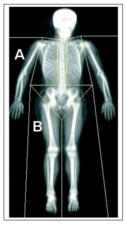
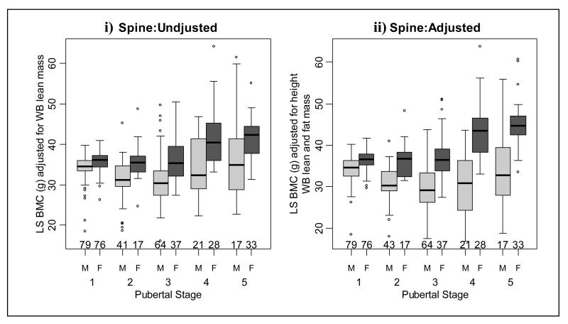
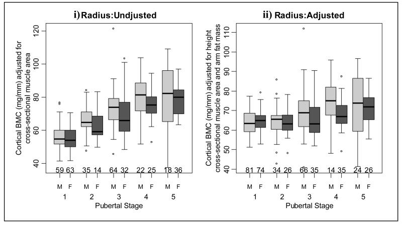
Methods: Four hundred forty-two children aged 5-18 years were studied. Measurements of bone mineral content (BMC), lean mass (LM) and fat mass of the whole body (WB), legs, arms and lumbar spine were obtained from dual-energy X-ray absorptiometry. Peripheral quantitative computed tomography was used to measure BMC of the radius diaphysis and cross-sectional muscle area (CSMA) of the mid-forearm. These measurements were used to describe differences between, and within, genders at each pubertal stage in BMC accrual relative to muscle, both before and after adjustment for height, regional fat and muscle at central and peripheral skeletal sites.
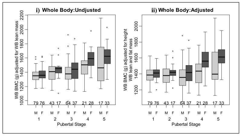
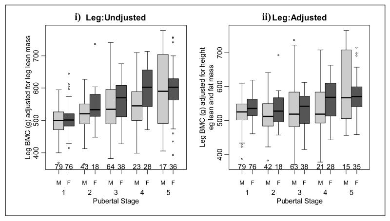
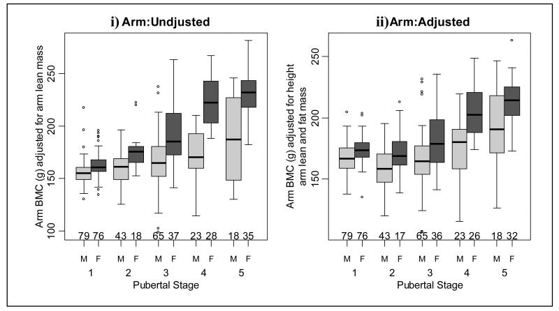
Results: In males, there were significant increases in adjusted WB and leg BMC at the end of pubertal development. Unadjusted and adjusted lumbar spine BMC increased at the onset of, and at the end, of puberty. Radius BMC increased at most pubertal stages. In females, there were increases in unadjusted and adjusted whole body BMC at late puberty, in leg BMC at the onset of puberty and at pubertal stage four. Unadjusted arm BMC increased at most pubertal stages; however, after adjustment, an increase occurred at pubertal stage four. Both adjusted and unadjusted lumbar spine BMC increased at pubertal stage four. Unadjusted radius BMC increased at most pubertal stages. Females had greater BMC at all skeletal sites, compared to males, except at the radius, where adjusted BMC was greater in males at pubertal stage four.
Conclusions: Males and females accrue more BMC in relation to lean mass at multiple skeletal sites as puberty proceeds. Females accrue more BMC in relation to lean mass, in comparison to males, at most skeletal sites.
Figures
Similar articles
-
Heterogeneity of growth of bone in children at the spine, radius and total skeleton.Growth Dev Aging. 1991 Winter;55(4):249-56. Growth Dev Aging. 1991. PMID: 1813443
-
Catch up in bone acquisition in young adult men with late normal puberty.J Bone Miner Res. 2012 Oct;27(10):2198-207. doi: 10.1002/jbmr.1675. J Bone Miner Res. 2012. PMID: 22653693
-
Bone geometry and density in the skeleton of pre-pubertal gymnasts and school children.Bone. 2005 Jun;36(6):1012-8. doi: 10.1016/j.bone.2005.03.001. Bone. 2005. PMID: 15876561
-
Effect of puberty on body composition.Curr Opin Endocrinol Diabetes Obes. 2009 Feb;16(1):10-5. doi: 10.1097/med.0b013e328320d54c. Curr Opin Endocrinol Diabetes Obes. 2009. PMID: 19115520 Review.
-
Puberty and body composition.Horm Res. 2003;60(Suppl 1):36-45. doi: 10.1159/000071224. Horm Res. 2003. PMID: 12955016 Review.
Cited by
-
Standardizing evaluation of pQCT image quality in the presence of subject movement: qualitative versus quantitative assessment.Calcif Tissue Int. 2014 Feb;94(2):202-11. doi: 10.1007/s00223-013-9803-x. Calcif Tissue Int. 2014. PMID: 24077875 Free PMC article.
-
Indian girls have higher bone mineral content per unit of lean body than boys through puberty.J Bone Miner Metab. 2018 May;36(3):364-371. doi: 10.1007/s00774-017-0843-6. Epub 2017 Jun 3. J Bone Miner Metab. 2018. PMID: 28580516
-
Bone Density in the Obese Child: Clinical Considerations and Diagnostic Challenges.Calcif Tissue Int. 2017 May;100(5):514-527. doi: 10.1007/s00223-016-0233-4. Epub 2017 Jan 20. Calcif Tissue Int. 2017. PMID: 28105511 Free PMC article.
-
Bone mineral content has stronger association with lean mass than fat mass among Indian urban adolescents.Indian J Endocrinol Metab. 2015 Sep-Oct;19(5):608-15. doi: 10.4103/2230-8210.163174. Indian J Endocrinol Metab. 2015. PMID: 26425468 Free PMC article.
-
Effects of reproduction on sexual dimorphisms in rat bone mechanics.J Biomech. 2018 Aug 22;77:40-47. doi: 10.1016/j.jbiomech.2018.06.023. Epub 2018 Jun 23. J Biomech. 2018. PMID: 29961584 Free PMC article.
References
-
- Parfitt AM. The two faces of growth: benefits and risks to bone integrity. Osteoporos Int. 1994;4:382–398. - PubMed
-
- Parfitt AM, Travers R, Rauch F, Glorieux FH. Structural and cellular changes during bone growth in healthy children. Bone. 2000;27:487–494. - PubMed
-
- Bradney M, Karlsson MK, Duan Y, Stuckey S, Bass S, Seeman E. Heterogeneity in the growth of the axial and appendicular skeleton in boys: Implications for the pathogenesis of bone fragility in men. J Bone Miner Res. 2000;15:1871–1878. - PubMed
-
- Lian JB, Stein GS, Canalis E, Gehron Robey P, Boskey AL. Bone formation: osteoblast lineage cells, growth factors, matrix proteins and the mineralization process. In: Favus MJ, editor. Primer on the metabolic bone diseases and disorders of mineral metabolism. Lippincott, Williams and Wilkins; Philadelphia: 1999. pp. 14–29.
Publication types
MeSH terms
Grants and funding
LinkOut - more resources
Full Text Sources
Medical
