Case Reports
. 2010 Jun;4(2):152-6.
doi: 10.1007/s12105-010-0172-8. Epub 2010 Mar 24.

Ectopic cervical thymoma: a report of two cases of a rare entity frequently misdiagnosed on fine needle aspiration cytology and frozen section

Affiliations
Case Reports

Ectopic cervical thymoma: a report of two cases of a rare entity frequently misdiagnosed on fine needle aspiration cytology and frozen section

Benedict Yan et al. Head Neck Pathol. 2010 Jun.

Abstract

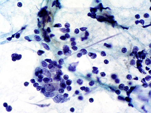
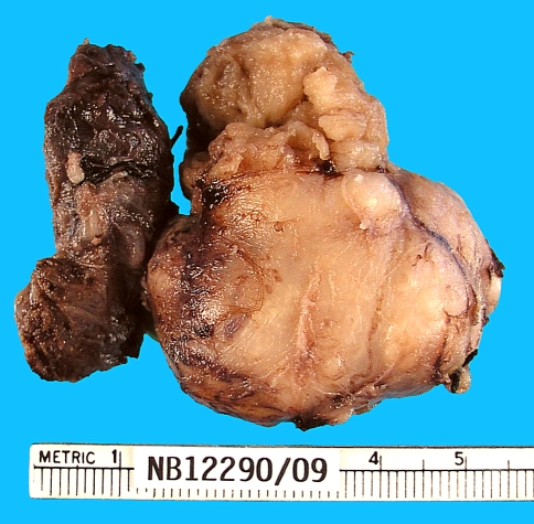
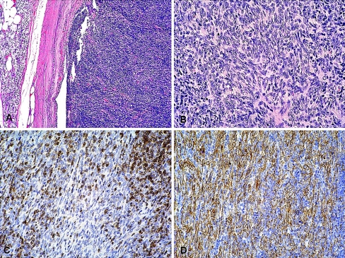
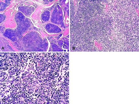
Ectopic cervical thymoma (ECT) is a rare tumor that is frequently misdiagnosed on fine needle aspiration cytology and frozen section. Using conventional light microscopy and immunohistochemistry, we characterized the only two cases of ECT found in our institutional files over a period of 20 years. Both tumors were classified as type AB-thymoma. Neoplastic cells expressed cytokeratins but not CD5. Non-neoplastic T-lymphocytes were positive for CD3 and CD5. Lymphocytes expressed CD1a in only one case. One tumor breached the capsule and had positive surgical margins. For this patient, adjuvant radiotherapy was given. The other patient has had an uneventful follow-up for 20 years with no other therapy than surgery. Both cases of ECT showed identical histomorphological and immunohistochemical features of type AB-thymomas originating in the thymus. Short follow-up precludes conclusion on the implication of positive margins in conjunction with adjuvant radiotherapy for one of the patients presented herein.


Figures

Fig. 1
Fig. 1
Case 1. Fine needle aspiration cytology showing a small lymphocytic population with a group of larger cells with oval nuclei and more abundant cytoplasm (×600)
Fig. 2
Fig. 2
Gross picture of a surgical specimen illustrating relation of tumor to the left thyroid lobe (Case 1). The thyroid is on the left side
Fig. 3
Fig. 3
Case 1. Photomicrographs (Hematoxylin and Eosin-stained sections). a Encapsulated tumor with a predominant lymphocyte-rich component. A rim of normal parathyroid tissue is present external to the capsule on the left (×100). b Lymphocyte-poor spindle cell area. The spindle cells show minimal cytologic atypia (×200). The Immunohistochemical study shows c positive reaction for CD1a in non-neoplastic lymphocytes and d positive reaction for cytokeratins (MNF116) in neoplastic cells (×200)
Fig. 4
Fig. 4
Case 2. Photomicrographs (Hematoxylin and Eosin-stained sections). a Lobulated, organoid tumor intersected by thick fibrous bands (×40). b Transition between lymphocyte-rich areas with predominantly spindle cell areas (×200). c Spindled to epithelioid neoplastic epithelial cells intimately admixed with lymphocytes (×400)

References

    1. Tunkel DE, Erozan YS, Weir EG. Ectopic cervical thymic tissue: diagnosis by fine needle aspiration. Arch Pathol Lab Med. 2001;125:278–281. - PubMed
    1. Oh YL, Ko YH, Ree HJ. Aspiration cytology of ectopic cervical thymoma mimicking a thyroid mass. A case report. Acta Cytol. 1998;42:1167–1171. - PubMed
    1. Chan JK, Rosai J. Tumors of the neck showing thymic or related branchial pouch differentiation: a unifying concept. Hum Pathol. 1991;22:349–367. doi: 10.1016/0046-8177(91)90083-2. - DOI - PubMed
    1. Gerhard R, Kanashiro EH, Kliemann Cm CM, et al. Fine-needle aspiration biopsy of ectopic cervical spindle-cell thymoma: a case report. Diagn Cytopathol. 2005;32:358–362. doi: 10.1002/dc.20258. - DOI - PubMed
    1. Chang ST, Chuang SS. Ectopic cervical thymoma: a mimic of T-lymphoblastic lymphoma. Pathol Res Pract. 2003;199:633–635. doi: 10.1078/0344-0338-00473. - DOI - PubMed

Publication types