Review
. 2010 Oct;16(5):467-91.
doi: 10.1089/ten.TEB.2009.0630.

Molecular regulation of contractile smooth muscle cell phenotype: implications for vascular tissue engineering

Affiliations
Review

Molecular regulation of contractile smooth muscle cell phenotype: implications for vascular tissue engineering

Jeffrey A Beamish et al. Tissue Eng Part B Rev. 2010 Oct.

Abstract

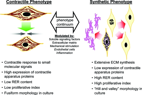
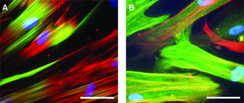
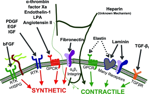
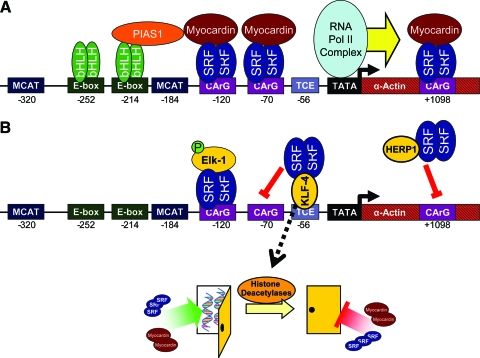
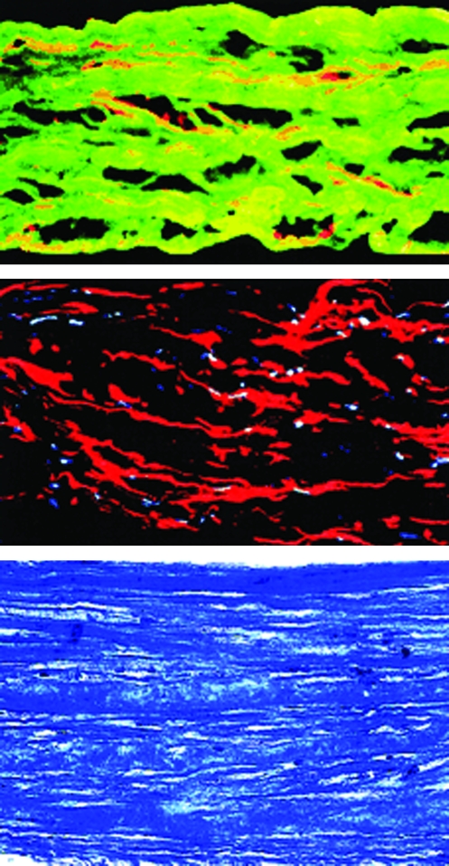
The molecular regulation of smooth muscle cell (SMC) behavior is reviewed, with particular emphasis on stimuli that promote the contractile phenotype. SMCs can shift reversibly along a continuum from a quiescent, contractile phenotype to a synthetic phenotype, which is characterized by proliferation and extracellular matrix (ECM) synthesis. This phenotypic plasticity can be harnessed for tissue engineering. Cultured synthetic SMCs have been used to engineer smooth muscle tissues with organized ECM and cell populations. However, returning SMCs to a contractile phenotype remains a key challenge. This review will integrate recent work on how soluble signaling factors, ECM, mechanical stimulation, and other cells contribute to the regulation of contractile SMC phenotype. The signal transduction pathways and mechanisms of gene expression induced by these stimuli are beginning to be elucidated and provide useful information for the quantitative analysis of SMC phenotype in engineered tissues. Progress in the development of tissue-engineered scaffold systems that implement biochemical, mechanical, or novel polymer fabrication approaches to promote contractile phenotype will also be reviewed. The application of an improved molecular understanding of SMC biology will facilitate the design of more potent cell-instructive scaffold systems to regulate SMC behavior.


Figures

FIG. 1.
FIG. 1.
Summary of characteristics of SMC phenotypes, which vary along a continuum from synthetic and proliferative to contractile and quiescent. The position along this continuum is modulated by a variety of extracellular signals. ECM, extracellular matrix; RER, rough endoplasmic reticulum; SMC, smooth muscle cell. Color images available online at www.liebertonline.com/ten.
FIG. 2.
FIG. 2.
Expression of contractile apparatus proteins in human coronary artery SMCs that have been cultured to re-induce contractile phenotype, observed by immunofluorescent staining. (A) Calponin (green) colocalizes (yellow) with SMαA (red) fibrils in the central region of the cells. (B) SM-22α (green) colocalizes (yellow) along the length of SMαA (red) fibrils. Variable staining between cells highlights the heterogeneity of cell populations along the contractile-synthetic phenotype continuum. Nuclei are counterstained with DAPI (blue). Scale bars: 50 μm. SMαA, smooth muscle α-actin. Color images available online at www.liebertonline.com/ten.
FIG. 3.
FIG. 3.
Brief overview of mechanisms involved in the modulation of SMC phenotype. The mechanism of action for heparin is unclear. Heparin may act by inhibiting binding of extracellular growth factors or secondary autocrine signaling factors, inhibiting intracellular signal transduction by these stimuli, and/or directly promoting contractile phenotype. Angiotensin II action can induce both synthetic and contractile characteristics. bFGF, basic fibroblast growth factor; PDGF, platelet-derived growth factors; EGF, epidermal growth factors; IGF, insulin-like growth factors; LPA, lysophosphatidic acid; TGF-β1, transforming growth factor beta 1; RTK, receptor tyrosine kinase; HSPG, heparan sulfate proteoglycan; GPCR, G-protein coupled receptor; TGFβR, TGF-β receptor. Color images available online at www.liebertonline.com/ten.
FIG. 4.
FIG. 4.
Molecular regulation of SMαA transcription, illustrating example mechanisms of transcriptional activation in differentiated SMCs (A) and mechanisms of downregulation (B). (A) Transcription is activated by SRF binding to CArG box up- and downstream of the TATA box, enhanced by the coactivator, myocardin. Additional elements further enhance transcription, such as bHLH transcription factors via PIAS-1. (B) Transcription is downregulated by phospho-Elk-1 blocking myocardin interactions with SRF. KLF-4 and HERP-1 block SRF binding to CArG boxes via sequestration. KLF-4 also activates histone deacetylases that close chromatin structure (represented as a closed door in the diagram), limiting transcription factor access to the promoter region. bHLH, basic helix-loop-helix; PIAS-1, protein inhibitor of activated STAT-1; SRF, serum response factor; KLF-4, Kruppel-like factor 4; HERP-1, Hairy- and enhancer of split-like-related repressor protein-1; TCE, TGF-β control element. Color images available online at www.liebertonline.com/ten.
FIG. 5.
FIG. 5.
Histology of a human TEVM (from Ref.). Top: Cross section of the TEVM immunolabeled for desmin (red) and type I collagen (green). Middle: Cross section of the TEVM immunolabeled for SMαA (red). Nuclei are stained blue. Bottom: Cross section of the TEVM stained with Masson's trichrome shows collagen in blue and cells in purple. TEVM, tissue-engineered vascular media. Color images available online at www.liebertonline.com/ten.

References

    1. Lloyd-Jones D. Adams R. Carnethon M. De Simone G. Ferguson T.B. Flegal K. Ford E. Furie K. Go A. Greenlund K. Haase N. Hailpern S. Ho M. Howard V. Kissela B. Kittner S. Lackland D. Lisabeth L. Marelli A. McDermott M. Meigs J. Mozaffarian D. Nichol G. O'Donnell C. Roger V. Rosamond W. Sacco R. Sorlie P. Stafford R. Steinberger J. Thom T. Wasserthiel-Smoller S. Wong N. Wylie-Rosett J. Hong Y. for the American Heart Association Statistics Committee and Stroke Statistics S. Heart disease and stroke statistics—2009 update: a report from the American Heart Association Statistics Committee and Stroke Statistics Subcommittee. Circulation. 2009;119:e21. - PubMed
    1. Laube H.R. Duwe J. Rutsch W. Konertz W. Clinical experience with autologous endothelial cell-seeded polytetrafluoroethylene coronary artery bypass grafts. J Thorac Cardiovasc Surg. 2000;120:134. - PubMed
    1. Greenwald S.E. Berry C.L. Improving vascular grafts: the importance of mechanical and haemodynamic properties. J Pathol. 2000;190:292. - PubMed
    1. Christen T. Verin V. Bochaton-Piallat M. Popowski Y. Ramaekers F. Debruyne P. Camenzind E. van Eys G. Gabbiani G. Mechanisms of neointima formation and remodeling in the porcine coronary artery. Circulation. 2001;103:882. - PubMed
    1. Sottiurai V.S. Yao J.S. Batson R.C. Sue S.L. Jones R. Nakamura Y.A. Distal anastomotic intimal hyperplasia: histopathologic character and biogenesis. Ann Vasc Surg. 1989;3:26. - PubMed

Publication types

LinkOut - more resources