. 2010 Apr;173(4):522-35.
doi: 10.1667/RR1882.1.

Early alterations in cytokine expression in adult compared to developing lung in mice after radiation exposure

Affiliations

Early alterations in cytokine expression in adult compared to developing lung in mice after radiation exposure

Carl J Johnston et al. Radiat Res. 2010 Apr.

Abstract

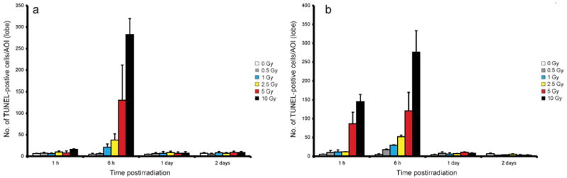
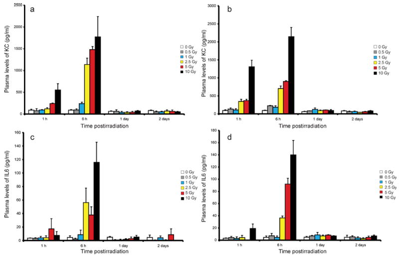
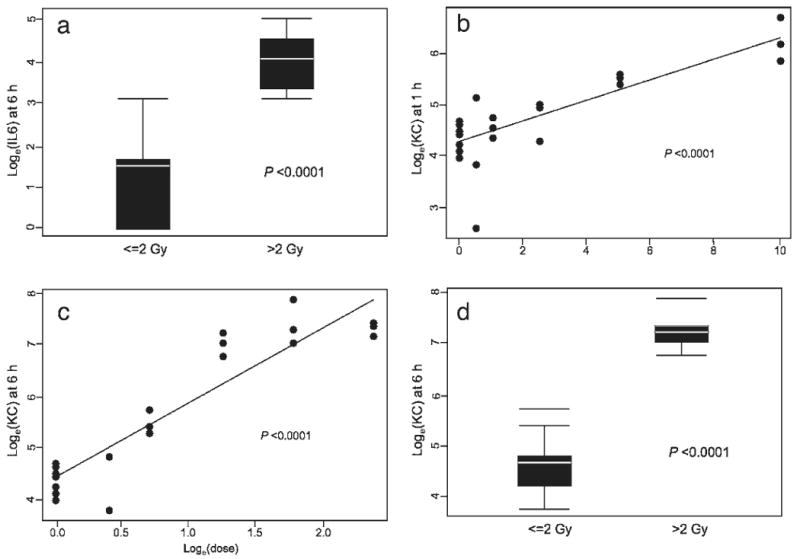
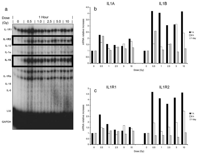
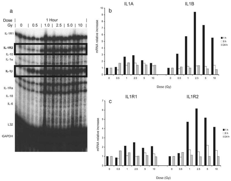
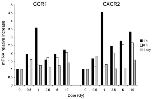
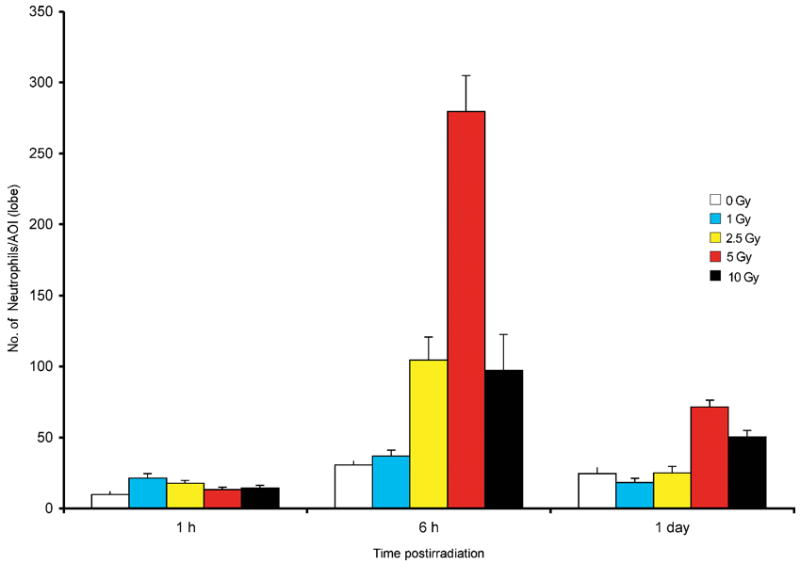
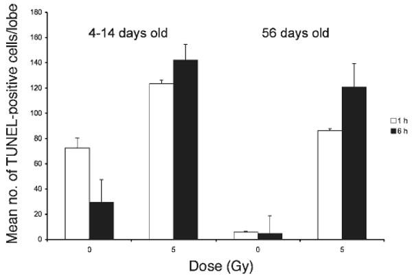
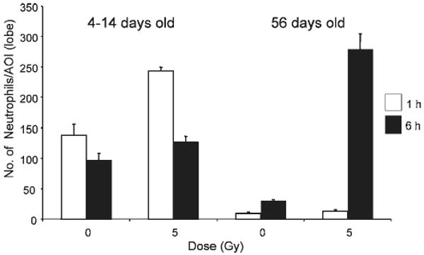
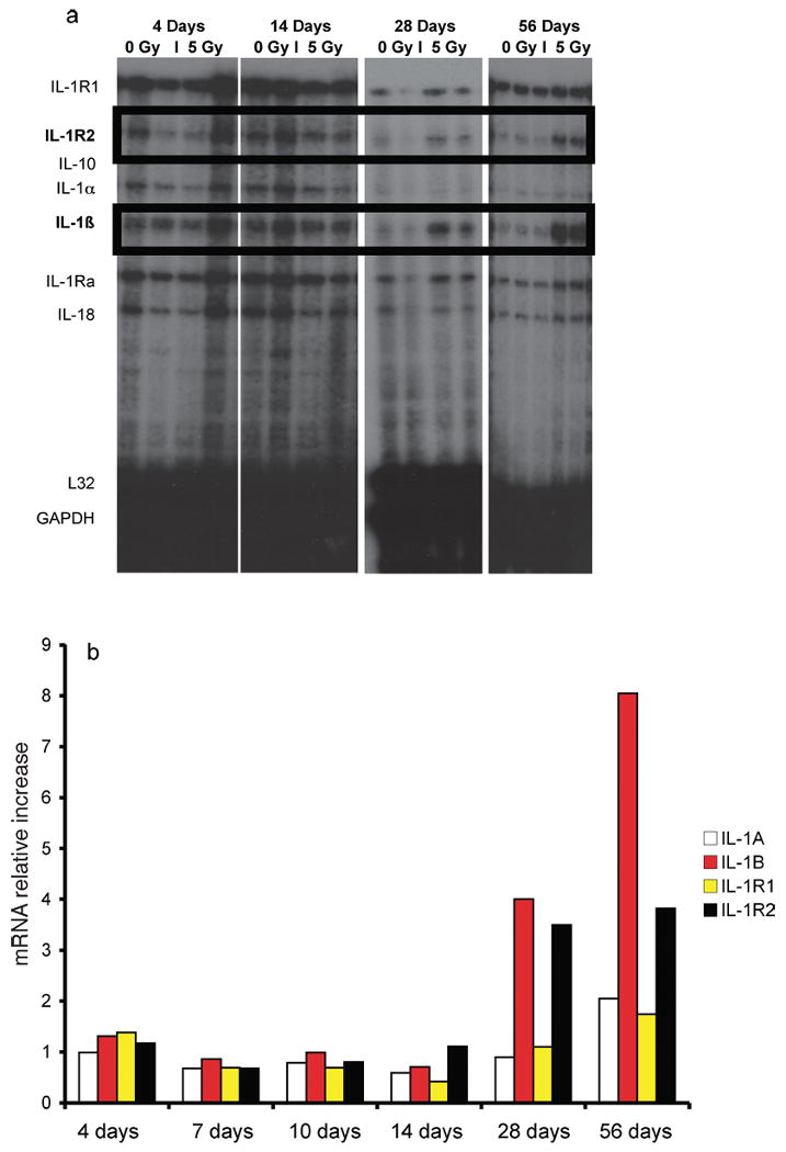
To assess early changes in the lung after low-dose radiation exposure that may serve as targets for mitigation of lung injury in the aftermath of a terrorist event, we analyzed cytokine expression after irradiation. Adult mice were studied after whole-lung or total-body irradiation. Mouse pups of different ages were also investigated after total-body irradiation. mRNA abundance was analyzed in tissue and plasma, and pathological changes were assessed. In lung tissue, dose-related changes were seen in IL1B, IL1R2 and CXCR2 mRNA expression at 1 and 6 h after irradiation, concurrent with increases in plasma protein levels of KC/CXCL1 and IL6. However, in the pups, changes in IL1 abundance were not detected until 28 days of age, coincident with the end of postnatal lung growth, although apoptosis was detected at all ages. In conclusion, although cytokines were expressed after low doses of radiation, their role in the progression of tissue response is yet to be determined. They may be candidates for use in marker-based biodosimetry. However, the lack of cytokine induction in early life suggests that different end points (and mitigating treatments) may be required for children.


Figures

FIG. 1
FIG. 1
Representative micrographs of TUNEL staining in C57BL/6J mouse lung after total-body irradiation with 1 Gy (panels a and b) and 10 Gy (panels c and d) at 1 and 6 h postirradiation, respectively.
FIG. 2
FIG. 2
TUNEL-positive cells in C57BL/6J mice at 1, 6, 24 or 48 h postirradiation after 0–10 Gy administered to the whole lung (panel a) or as total-body irradiation (panel b). n = 10 per group. Error limits are standard errors.
FIG. 3
FIG. 3
Analysis of circulating KC/CXCL1 (panels a and b) and IL6 (panels c and d) after 0–10 Gy whole-lung and total-body irradiation, respectively, of C57BL/6J mice at 1, 6, 24 or 48 h. n = 10 per group.
FIG. 4
FIG. 4
Statistical analysis to assess the significance of cytokine expression, as measured by multiplex assay, in the plasma after whole-lung irradiation: plasma levels of IL6 at 6 h postirradiation (panel a) and KC/CXCL1 at 1 h (panel b) and 6 h (panels c and d) postirradiation, respectively. Error limits are standard errors.
FIG. 5
FIG. 5
Statistical analysis to assess the significance of the expression, as measured by multiplex assay, of the plasma levels of IL6 and KC/CXCL1 in C57BL/6J mice after whole-lung (panels a and d) or total-body irradiation (TBI) (panels b and c) at 6 h postirradiation. Error limits are standard errors.
FIG. 6
FIG. 6
Panel a: RNase protection assay measuring interleukin mRNA abundance in the lungs after whole-lung irradiation. IL1R2 and IL1B are highlighted to aid in interpretation. Time course of changes in the relative mRNA abundance of (panel b) IL1A and IL1B and (panel c) IL1R1 and IL1R2 in lungs after irradiation. RNase protection assays were quantified by PhosphorImager analysis. All values were normalized to constitutively expressed mRNA encoding L32. Each bar represents the mean for a minimum of five mice. Error limits are standard errors.
FIG. 7
FIG. 7
Panel a: RNase protection assay measuring interleukin mRNA abundance in the lungs 1 h after total-body irradiation at 1 h. IL1R2 and IL1B are highlighted to aid in interpretation. Time course of changes in relative mRNA abundance of (panel b) IL1A and IL1B and (panel c) IL1R1 and IL1R2 in lungs after irradiation. RNase protection assays were quantified by PhosphorImager analysis. All values were normalized to constitutively expressed mRNA encoding L32. Each bar represents the mean for a minimum of five mice.
FIG. 8
FIG. 8
Time course of changes in relative mRNA abundance of CCR1 and CXCR2 after whole-lung irradiation. RNase protection assays were quantified by PhosphorImager analysis. All values were normalized to constitutively expressed mRNA encoding L32. Each bar represents the mean of a minimum of five mice.
FIG. 9
FIG. 9
Number of neutrophils in the lungs of C57BL/6J mice after whole-lung irradiation. Error limits are standard errors.
FIG. 10
FIG. 10
TUNEL-positive cells in mouse pups (4–14 days old) and adult (56 days old) C57BL/6J after total-body irradiation. Error limits are standard errors.
FIG. 11
FIG. 11
Circulating KC/CXCL1 after 0 or 5 Gy total-body irradiation of mouse pups (4–14 days old) and adult (56 days old) at 6 h postirradiation. Error limits are standard errors.
FIG. 12
FIG. 12
Number of neutrophils in the lungs of mouse pups (4–14 days old) and adults (56 days old) after 0 or 5 Gy total-lung irradiation. Error limits are standard errors.
FIG. 13
FIG. 13
Panel a: RNase protection assay measuring interleukin mRNA abundance in the lungs of adult (56 days old) mice and mouse pups 1 h after 0 or 5 Gy total-body irradiation. RNase protection assay: 10 μg total lung RNA hybridized with 5.2 × 10⁵ cpm for 18 h at 56°C. Panel b: Time course of changes in relative mRNA abundance of IL1A, IL1B, IL1R1 and IL1R2 in lungs of adult (56 days old) C57BL/6J mice and mouse pups at 1 h postirradiation. RNase protection assays were quantified by PhosphorImager analysis. All values were normalized to constitutively expressed mRNA encoding L32. Each bar represents the mean of a minimum of three mice.

Similar articles

Cited by

References

    1. Gonzalez AJ, Lauriston S. Taylor Lecture: Radiation protection in the aftermath of a terrorist attack involving exposure to ionizing radiation. Health Phys. 2005;89:418–446. - PubMed
    1. NIH Publication No 05-5608. U.S. Department of Health and Human Services; Washington, DC: 2005. NIH Strategic Plan and Research Agenda for Medical Countermeasures Against Radiological and Nuclear Threats.
    1. Continuation of national emergency regarding the proliferation of weapons of mass destruction. Fed Regist. 2006;71:64109.
    1. UNSCEAR. Report to the General Assembly, Annex J: Exposures and Effects of the Chernobyl Accident. United Nations; New York: 2000. Sources, Effects and Risks of Ionizing Radiation.
    1. Hirama T, Tanosaki S, Kandatsu S, Kuroiwa N, Kamada T, Tsuji H, Yamada S, Katoh H, Yamamoto N, Akashi M. Initial medical management of patients severely irradiated in the Tokai-mura criticality accident. Br J Radiol. 2003;76:246–253. - PubMed

Publication types