. 2010 Mar 24:8:19.
doi: 10.1186/1741-7015-8-19.

The cytoprotective drug amifostine modifies both expression and activity of the pro-angiogenic factor VEGF-A

Affiliations

The cytoprotective drug amifostine modifies both expression and activity of the pro-angiogenic factor VEGF-A

S Dedieu et al. BMC Med. .

Abstract

Background: Amifostine (WR-2721, delivered as Ethyol) is a phosphorylated aminothiol compound clinically used in addition to cis-platinum to reduce the toxic side effects of therapeutic treatment on normal cells without reducing their efficacy on tumour cells. Its mechanism of action is attributed to the free radical scavenging properties of its active dephosphorylated metabolite WR-1065. However, amifostine has also been described as a potent hypoxia-mimetic compound and as a strong p53 inducer; both effects are known to potently modulate vascular endothelial growth factor (VEGF-A) expression. The angiogenic properties of this drug have not been clearly defined.

Methods: Cancer cell lines and endothelial cells were used in culture and treated with Amifostine in order to study (i) the expression of angiogenesis related genes and proteins and (ii) the effects of the drug on VEGF-A induced in vitro angiogenesis.

Results: We demonstrated that the treatment of several human cancer cell lines with therapeutical doses of WR-1065 led to a strong induction of different VEGF-A mRNA isoforms independently of HIF-1alpha. VEGF-A induction by WR-1065 depends on the activation of the eIF2alpha/ATF4 pathway. This up-regulation of VEGF-A mRNA was accompanied by an increased secretion of VEGF-A proteins fully active in stimulating vascular endothelial cells (EC). Nevertheless, direct treatment of EC with amifostine impaired their ability to respond to exogenous VEGF-A, an effect that correlated to the down-regulation of VEGFR-2 expression, to the reduction in cell surface binding of VEGF-A and to the decreased phosphorylation of the downstream p42/44 kinases.

Conclusions: Taken together, our results indicate that amifostine treatment modulates tumour angiogenesis by two apparently opposite mechanisms - the increased VEGF-A expression by tumour cells and the inhibition of EC capacity to respond to VEGF-A stimulation.


Figures
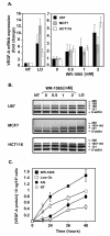
Figure 1
Figure 1
Amifostine enhances vascular endothelial growth factor A (VEGF-A) mRNA and protein expression in cancer cells. MCF7 and HCT116 carcinoma cells, as well as U87 glioma cells, were grown up to 70% confluence in 10-cm culture dishes. Cells were then incubated in freshly added complete medium under classical conditions of oxygen (NT, 20% 02) or low oxygen conditions (LO, low O2, 3% 02), or in the presence of the indicated concentrations of WR-1065 under 20% of oxygen. (A) Amifostine increases VEGF-A mRNA levels in U87, MCF7 and HCT116 cells. Following a 16-h (MCF7 cells, grey bars) or a 24-h (HCT116 cells, white bars; U87 cells, black bars) incubation time, total mRNA was isolated. Expression of VEGF-A mRNA in cells was measured by quantitative polymerase chain reaction using primers amplifying all the VEGF-A isoforms. Histogram values represent the level of expression of all VEGF-A splice variants, normalized to α-tubulin. Results are the mean values ± standard error of mean of three independent experiments. (B) Amifostine increases specific VEGF-A mRNA isoforms. Shown is a representative gel electrophoresis pattern of the different VEGF-A splice variants in U87, MCF7 and HCT116 cells, β-actin being used as standard. (C) Amifostine increases VEGF-A protein secretion by MCF7 cells. Cells were treated up to 2 days in complete medium, and conditioned media were collected after 24 h, 36 h and 48 h of treatment. VEGF-A protein secretion was measured by ELISA using supernatants of cells grown under low levels of oxygen (3% O2,) or 20% of oxygen, and of cells treated with 1 mM WR-1065 in the presence of aminoguanidine (AG) or with AG alone. Results shown are representative of three independent experiments done in triplicates.
Figure 2
Figure 2
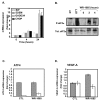
Amifostine upregulates vascular endothelial growth factor A (VEGF-A) mRNA production independently of hypoxia-inducible factor (HIF). MCF7 cells were incubated in complete medium with WR-1065 in the presence of aminoguanidine (AG) or were subjected to low levels of oxygen (3% 02,). (A) Amifostine treatment does not lead to any accumulation of HIF proteins. Cells were treated for 6 h under 20% of oxygen (20% O2) with or without 1 mM WR-1065; or low oxygen conditions (3% O2). HIF-1α and HIF-2α were detected by Western blot (Ku80 used as internal control). (B) Amifostine treatment does not lead to the induction of HIF-1 target genes. Cells were treated with WR-1065, in the presence of AG, for 16 h or exposed to low oxygen conditions (3% O2) for 16 h. Total mRNA were isolated and the expression of HK-2 and GLUT-1 genes were assessed by quantitative polymerase chain reaction Q-PCR); β-actin served as internal control. (C/D) Amifostine-mediated VEGF-A upregulation is not impaired by a HIF-1α.shRNA (small hairpin RNA). (C) Western blot analysis of HIF-1α protein levels in MCF7 cells transduced with RFP.shRNA (control) and HIF-1α.shRNA constructions. Cells were treated with 1 mM WR-1065 for 16 h (upper panel, "+"), or exposed to low oxygen conditions (3% O2) or classical conditions (20% O2) for 6 h (lower panel). Proteins were analysed by Western blot using specific antibodies for HIF-1α and for Ku80 (control of loading). (D) VEGF-A mRNA expression was assessed by Q-PCR in MCF7 cells transduced with lentiviral constructs expressing either HIF-1α.shRNA or control RFP.shRNA (grey and white bars, respectively). Cells were incubated for 16 h in the presence of WR-1065 or subjected to low oxygen conditions (3% O2). Expression of global VEGF-A mRNA in HIF-1α.shRNA-expressing cells was determined by Q-PCR and expressed in fold-change relative to untransduced-cells left untreated. Significant expression changes were determined by Student's paired tests.
Figure 3
Figure 3
Amifostine activates the unfolded protein response-dependent signalling pathways. MCF7 and U87 cells were treated for increasing periods of time with 2 mM WR-1065 or 10 μg/mL Tunicamycin (Tun) in complete culture medium. Proteins and mRNA were collected at several time points, and protein and gene expression were assessed by Western-Blot and polymerase chain reaction (PCR) analyses. (A) Amifostine differentially up-regulates BIP, EDEM, GADD34 and CHOP genes in MCF7 cells. Quantitative-PCR profiles of BIP, EDEM, GADD34 and CHOP genes, in MCF7 cells treated with WR-1065. Results were normalized using α-tubulin mRNA and expressed as ratios between the treated [aminoguanidine (AG)+WR1065] and untreated (AG alone) conditions. The time zero of the experiment is set to 1. (B) Amifostine triggers eIF2α phosphorylation in MCF7 cells. Western-blot analysis of total (Tot.eIF2α) and phosphorylated eIF2α (P-eIF2α) in MCF7 cells treated or not with 2 mM WR-1065. A 1 h treatment with 2 mM dithiothreitol was used as a positive control for eIF2α phosphorylation. Total protein extracts were resolved on 12% SDS-PAGE, and protein levels were assessed using specific anti-eIF2α and anti-P-eIF2α antibodies. (C/D) Amifostine-induced vascular endothelial growth factor A (VEGF-A) up-regulation requires activating transcription factor 4 (ATF4). MCF7 cells were transduced with a small interfering (si)RNA directed against ATF4 (SiATF4, white bars) or with the same siRNA mutated in three nucleotides (SiMUT, grey bars), used as control;. Transduced cells were treated for 24 h with AG alone (CTL) or in combination with 2 mM WR-1065. (C) ATF4 gene expression was assessed by real-time reverse polymerase chain reaction (RT-PCR). (D) VEGF-A gene expression was assessed by real-time RT-PCR. ATF4 and VEGF-A mRNA expression levels in amifostine-treated cells are expressed as fold changes to the control condition (AG alone), which were set to 1 for SiATF4 and SiMUT. Error bars correspond to standard deviations for each triplicate determination.
Figure 4
Figure 4
Amifostine induces a paracrine stimulatory effect on endothelial cells (ECs). MCF7 cells were grown to subconfluence and incubated for 48 h in the presence or absence of 1 mM WR-1065. Conditioned media (CM) were then collected and dialyzed to remove WR-1065. Cells were counted. Vascular endothelial growth factor A (VEGF-A) neutralizing antibody (1 μg/mL) were added or not to CM from WR-1065-treated (WR-1065) or AG-treated control cells (CTL). These control cells were supplemented (CTL+VEGF) or not (CTL) with 10 ng/mL VEGF-A. CM were added to HUVEC that have previously been starved in fetal bovine serum-free Dulbecco's modified eagle medium (see Materials and Methods section). Human umbilcal vein EC metabolic activity was measured using the Wst1 assay over a 14 h incubation time in the presence of CM. Results are expressed as means ± standard deviation of the OD (Optical Density) measured at 440 nm from six independent cell culture wells. Significant changes in OD, as determined by Student paired tests, are depicted on the histogram (* P < 0.05; ** P < 0.01).
Figure 5
Figure 5
Amifostine inhibits human umbilcal vein endothelial cells (HUVEC) proliferation and vascular endothelial growth factor A (VEGF-A)-induced migration and differentiation. (A) Amifostine inhibits HUVEC proliferation. HUVEC growing in endothelial growth medium-2 supplemented with 0, 0.25, 0.5, 1 or 2 mM of WR-2721 were counted for 3 days. The insert shows the inhibition of cell proliferation after 2 days of treatment, in percentage of untreated cells. (B) Amifostine inhibits VEGF-A dependant HUVEC migration. A Transwell migration assay was used. HUVEC were seeded in upper compartments and incubated in Dulbecco's modified eagle medium containing (DMEM) 0.5% fetal bovine serum (FBS), 0.1% bovine serum albumine and increasing concentrations of WR-1065. 10 ng/mL VEGF-A were, or were not, added in the lower compartment and cells were allowed to migrate for 7 h. Cells that migrated were fixed, stained and quantified. Left panel: quantification of VEGF-A-dependent HUVEC migration (% of the migration versus control untreated cells). Significant changes were determined by Student paired tests (*P < 0.05; **P < 0.01). Right panel: corresponding photomicrographs of HUVEC. (C) Amifostine inhibits VEGF-A induced capillary-like structures formation. HUVEC were seeded on GFR-Matrigel. After a 1 h incubation in 0.5% FBS-containing DMEM to allow cell adhesion, cells were treated with WR-1065 for 3 h, in the presence or absence of 10 ng/mL VEGF-A. Total tubule length was determined using NIS image analysis software. Left panel: quantification of total tubule length in the different experimental conditions. Results are expressed as percentages of the average tubule length in the control condition, set to 100%. The control condition corresponds to absence of both VEGF-A and WR-1065. Significant changes, were determined by Student paired tests (*P < 0.05; **P < 0.01). Right panel: phase-contrast microphotographs of tubule networks formed by HUVEC, at a 200-fold magnification.
Figure 6
Figure 6
Amifostine inhibits vascular endothelial growth factor (VEGF)R2 expression, VEGF-A binding and signalling in human umbilcal vein endothelial cells (HUVEC). A Amifostine treatment inhibits VEGFR-2 protein expression in HUVEC. After a 24 h-starvation, subconfluent HUVEC were incubated in the presence or absence of 1 mM WR-1065 for indicated period of time and total proteins were collected in an RIPA buffer and resolved on a 7.5% acrylamide SDS-PAGE gel. Proteins were blotted onto a nitrocellulose membrane and probed with a mouse monoclonal antibody against VEGFR-2. Equal loading of proteins was confirmed by β-actin detection. VEGFR-2 migrated at gel positions corresponding to the 150- and 200-kDa forms, as already reported in HUVEC. The blot is representative of three independent experiments. (B) Amifostine treatment impairs VEGF-A binding on HUVEC. HUVEC monolayers (7000 cells/cm2) were allowed to grow for 3 days and then incubated for 12 h in the absence or presence of 1 mM WR-1065. Cells were then exposed to increasing concentrations of 125I-labelled VEGF-A for 2 h at 4°C. The amount of specifically bound iodinated VEGF-A was then determined. Insert represents Scatchard plot corresponding to the saturation curves. The experiment was carried out three times with similar results. (C) Amifostine treatment impairs mitogen activated protein kiinase signalling in HUVEC. Subconfluent HUVEC in serum-free Dulbecco's modified eagle medium were treated with 10 ng/mL VEGF-A for 10 min, alone or together with 1 mM WR-1065. Cytoplasmic proteins were then collected and were resolved on 15% SDS-PAGE. Total (phosphorylated and unphosphorylated) p42/p44 proteins as well as the phosphorylated proteins (P-p42/P-p44) were probed using specific antibodies. Signal was assessed by quantification of chemoluminescence, and ratios between signal intensities of P-p42/P-p44 and total p42/p44 are given beneath the gel. They were normalized to the ratio obtained for untreated cells (VEGF -, WR-1065-), which is set to 1.

Similar articles

Cited by

References

    1. News and products notes. Formulary. 1996;31:152.
    1. Shaw LM, Bonner HS, Brown DQ. Metabolic pathways of WR-2721 (ethyol, amifostine) in the BALB/c mouse. Drug Metab Dispos. 1994;22:895–902. - PubMed
    1. Calabro-Jones PM, Fahey RC, Smoluk GD, Ward JF. Alkaline phosphatase promotes radioprotection and accumulation of WR-1065 in V79-171 cells incubated in medium containing WR-2721. Int J Radiat Biol Relat Stud Phys Chem Med. 1985;47:23–27. doi: 10.1080/09553008514550041. - DOI - PubMed
    1. Koukourakis MI. Amifostine in clinical oncology: current use and future applications. Anticancer Drugs. 2002;13:181–209. doi: 10.1097/00001813-200203000-00001. - DOI - PubMed
    1. Smoluk GD, Fahey RC, Calabro-Jones PM, Aguilera JA, Ward JF. Radioprotection of cells in culture by WR-2721 and derivatives: form of the drug responsible for protection. Cancer Res. 1988;48:3641–3647. - PubMed

Publication types

MeSH terms

Substances