Comparative Study
. 2010 Jun 5;401(2):266-79.
doi: 10.1016/j.virol.2010.02.027. Epub 2010 Mar 24.

Myxoma and vaccinia viruses exploit different mechanisms to enter and infect human cancer cells

Affiliations
Comparative Study

Myxoma and vaccinia viruses exploit different mechanisms to enter and infect human cancer cells

Nancy Y Villa et al. Virology. .

Abstract

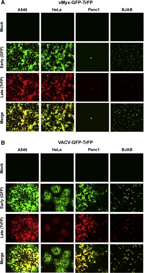
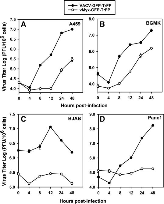
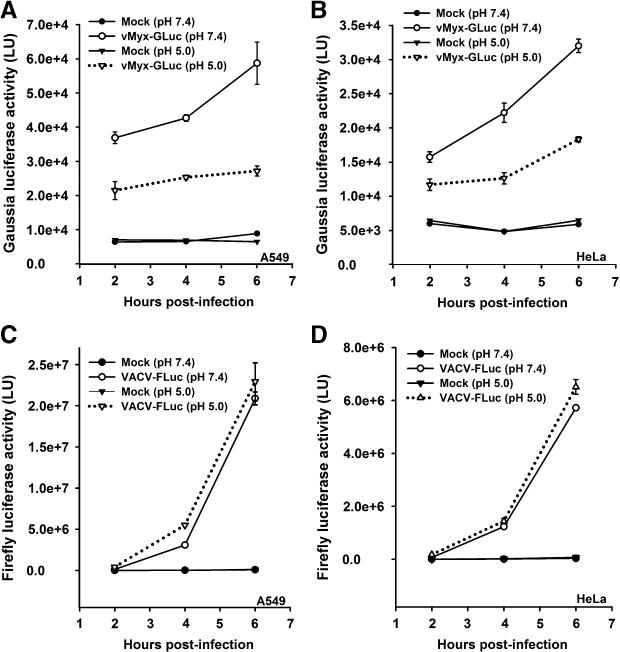
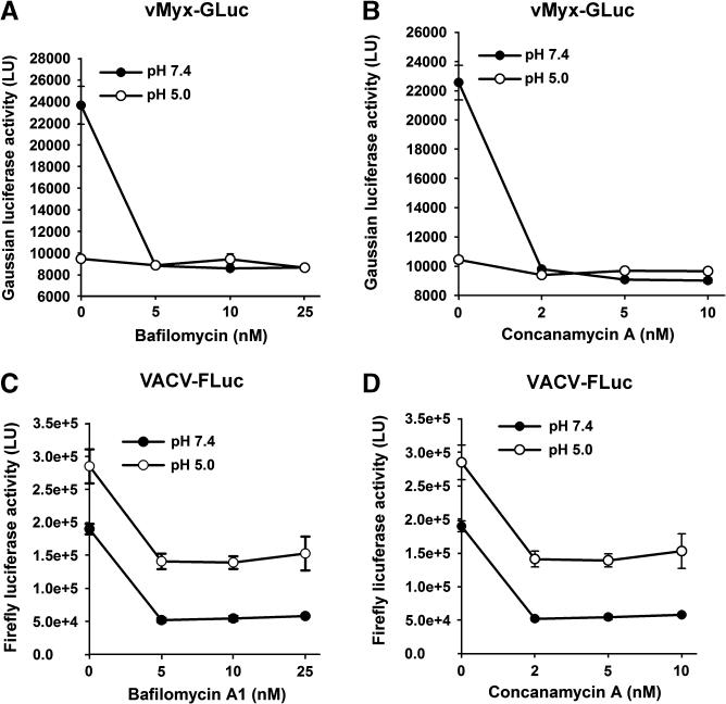
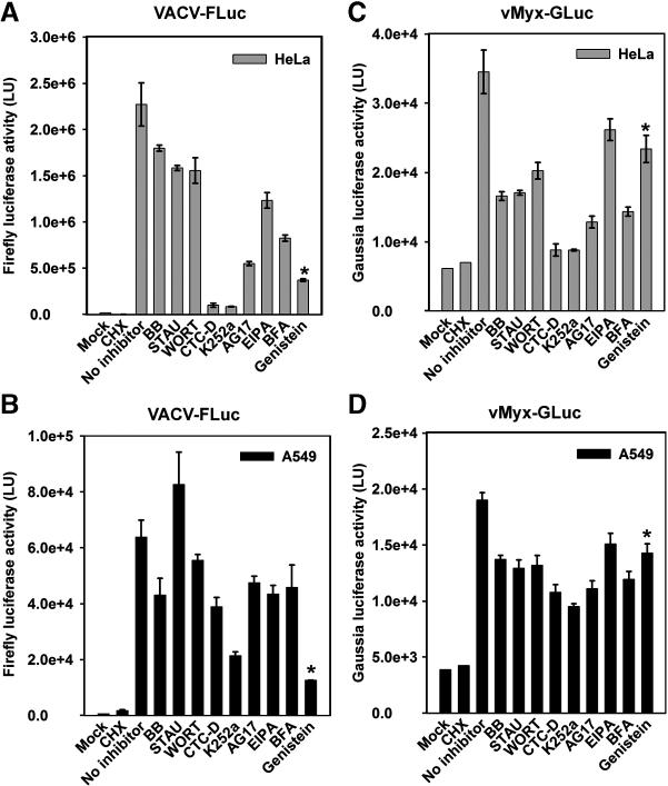
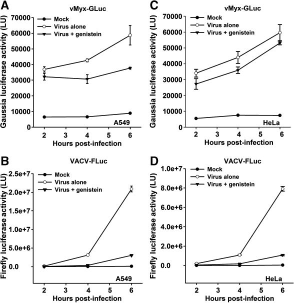
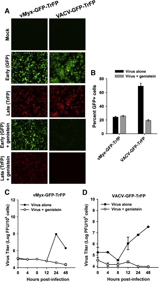
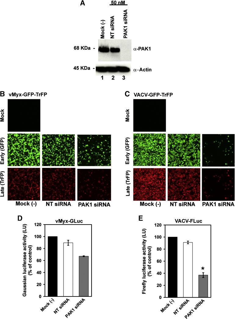
Myxoma (MYXV) and vaccinia (VACV) viruses have recently emerged as potential oncolytic agents that can infect and kill different human cancer cells. Although both are structurally similar, it is unknown whether the pathway(s) used by these poxviruses to enter and cause oncolysis in cancer cells are mechanistically similar. Here, we compared the entry of MYXV and VACV-WR into various human cancer cells and observed significant differences: 1--low-pH treatment accelerates fusion-mediated entry of VACV but not MYXV, 2--the tyrosine kinase inhibitor genistein inhibits entry of VACV, but not MYXV, 3--knockdown of PAK1 revealed that it is required for a late stage event downstream of MYXV entry into cancer cells, whereas PAK1 is required for VACV entry into the same target cells. These results suggest that VACV and MYXV exploit different mechanisms to enter into human cancer cells, thus providing some rationale for their divergent cancer cell tropisms.


Figures

Fig. 1
Fig. 1
MYXV and VACV replication and spread is cell line-dependent. Selected human cancer cell lines were infected with purified MV of (A) vMyx-GFP-TrFP, or (B) VACV-GFP-TrFP MVs, at a multiplicity of 0.1 (MOI 0.1). At 48 hour after infection the formation of foci (for MYXV) or plaques (for VACV) was visualized by fluorescence microscopy.
Fig. 2
Fig. 2
One-step growth of MYXV and VACV is cell line-specific. Single growth curves were generated to evaluate the replication of MYXV and VACV in (A) A549, (B) BGMK (C) BJAB and (D) Panc1 cells. Cells were infected with either vMyx-GFP-TrFP or VACV-GFP-TrFP at a multiplicity of 5.0. Titers are expressed as log PFU/106 cells. The single growth curve for BGMK was used as a positive control, since this cell line is optimally permissive for both viruses.
Fig. 3
Fig. 3
VACV and MYXV entry into cancer cells is differentially affected by brief low-pH exposure. A549 cells (panels A and C) and HeLa cells (panels B and D) were infected with vMyx-GLuc (panels A and B) or VACV-FLuc (panels C and D) at MOI of 5.0. After virus adsorption to the cells, unbound virus was removed and cells were washed with PBS followed by 3 min treatment in neutral pH (7.4) or low pH (5.0) at 37°C. Cells were washed in neutral pH media and the infection was allowed to proceed for different time points (2, 4, and 6 hours). At the given time point, cells were assayed for luciferase activity. Data represent the average ± standard error of samples in triplicate. Luciferase activity is reported in light units (LU).
Fig. 4
Fig. 4
Inhibitors of endosomal acidification decrease both VACV and MYXV entry. HeLa cells were pretreated with the indicated inhibitor for 1 hour at 37°C. While VACV-FLuc (panels C and D) was adsorbed to the cells at 4°C for 1 hour, vMyx-GLuc (panels A and B) was adsorbed to the cells at 37°C for 1 hour. Un-adsorbed viruses were removed and cells treated with either neutral pH (7.4), or acidic pH (5.0) in identical fashion as described elsewhere in this manuscript. Two hours after infection, cells were assayed for luciferase activity. Data represent the average of triplicates ± standard error.
Fig. 5
Fig. 5
MYXV and VACV entry is affected differentially by drug inhibitors. Prior to infection, cells HeLa (panels A and C) and A549 (panels B and D) were treated with each indicated inhibitor at the appropriate concentration during 1 hour at 37°C. Either vMyx-GLuc (panels C and D), or VACV-FLuc (panels A and B) was adsorbed to the cells at 37°C for 1 hour in the presence of inhibitor. After adsorption, cells were washed and then incubated with media supplemented with the appropriate inhibitor for 1 hour. Cells were subsequently assayed for luciferase activity. Cycloheximide (CHX) was added to the cells to prevent the early viral protein synthesis and serves as an internal control to quantify newly synthesized luciferase. Data represent the average ± standard error of samples in triplicate, and are expressed in light units (LU).
Fig.6
Fig.6
Genistein differentially affects MYXV and VACV entry in a time-independent manner. Kinetic experiments were performed for A549 (panels A and B) and HeLa (panels C and D). Cells pre-treated with genistein were infected with vMyx-GLuc (panels A and C) or VACV-FLuc (panels B and D) for 1 hour, at 37°C. At the given time points post-infection, luciferase activity is expressed in light units, (LU).
Fig. 7
Fig. 7
Genistein differentially affects MYXV and VACV. (A) HeLa cells were treated with genistein for 1 hour and then infected with vMyx-GFP-TrFP or VACV-GFP-TrFP at a MOI of 1.0. After 1 hour of virus adsorption, cells were incubated in a media containing genistein. Virus propagation was assessed by fluorescence microscopy 72 hours post-infection. (B) To measure cell-cell spread, the percentage of GFP+ cells in untreated (no genistein) and genistein-treated samples was determined by flow cytometry. Cells were infected with each virus at a MOI of 1.0, trypsinized and fixed in formaldehyde before analysis. (C) and (D): To investigate the effect of genistein on MYXV and VACV virus titers, cells were harvested at the given time points and then lysed by repeated freeze-thawing. Virus titers were determined as described in Materials and Methods.
Fig. 8
Fig. 8
Entry of MYXV and VACV into cancer cells is differentially affected by knockdown of PAK1. (A) Representative Western blots confirming PAK1 knock down. HeLa cells were transfected for 72 hours with 50 nM siRNA directed against PAK1 (lane 3), or against a non-targeting siRNA, (NT siRNA), (lane 2). Untransfected cells are referred as mock (-) (lane 1). Equal sample loading was confirmed by detection of the housekeeping protein actin. Replication of vMyx-GFP-TrFP (panel B) and VACV-GFP-TrFP (panel C) in HeLa cells was evaluated by fluorescence microscopy. 72 hours after transfection, cells were infected with the recombinant fluorescent viruses at a MOI of 1.0. Formation of MYXV foci, or VACV plaques was evaluated 48 hpi. Entry of MYXV-GLuc (D) or VACV-FLuc (E) into cells was assessed 72 hours post-transfection by measuring luciferase activity after 2 hours infection with vMyx-GLuc or VACV-FLuc at MOI of 5.0.

Similar articles

Cited by

References

    1. Andres A, Donovan SM, Kuhlenschmidt MS. Soy isoflavones and virus infections. J. Nutr, Biochem. 2009;20:563–569. - PMC - PubMed
    1. Andres A, Donovan SM, Kuhlenschmidt TB, Kuhlenschmidt MS. Isoflavones at concentrations present in soy infant formula inhibit rotavirus infection in vitro. J Nutr. 2007;137:2068–73. - PubMed
    1. Barrett JW, Alston LR, Wang F, Stanford MM, Gilbert PA, Gao X, Jimenez J, Villeneuve D, Forsyth P, McFadden G. Identification of host range mutants of myxoma virus with altered oncolytic potential in human glioma cells. J. Neurovirol. 2007a;13:549–560. - PubMed
    1. Barrett JW, Sypula J, Wang F, Alston LR, Shao Z, Gao X, Irvine TS, McFadden G. Myxoma virus M063R is a host range gene essential for virus replication in rabbit cells. Virology. 2007b;361:123–132. - PubMed
    1. Bartee E, Mohamed RM, Lopez MC, Baker HV, McFadden G. The Addition of Tumor Necrosis Factor plus Beta Interferon Induces a Novel Synergistic Antiviral State against Poxviruses in Primary Human Fibroblasts. J. Virol. 2009;83:498–511. - PMC - PubMed

Publication types