. 2010 May 21;285(21):16360-8.
doi: 10.1074/jbc.M110.100669. Epub 2010 Mar 24.

The Salmonella type III secretion effector, salmonella leucine-rich repeat protein (SlrP), targets the human chaperone ERdj3

Affiliations

The Salmonella type III secretion effector, salmonella leucine-rich repeat protein (SlrP), targets the human chaperone ERdj3

Joaquín Bernal-Bayard et al. J Biol Chem. .

Abstract

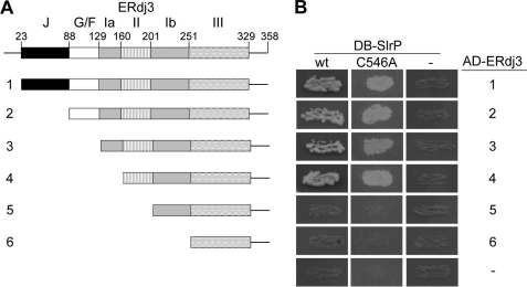
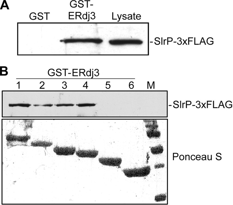
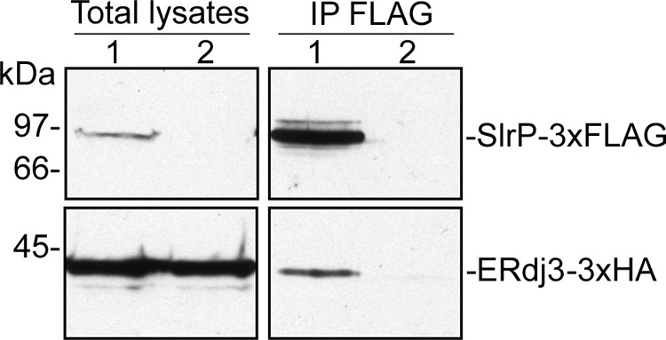
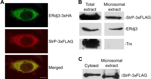
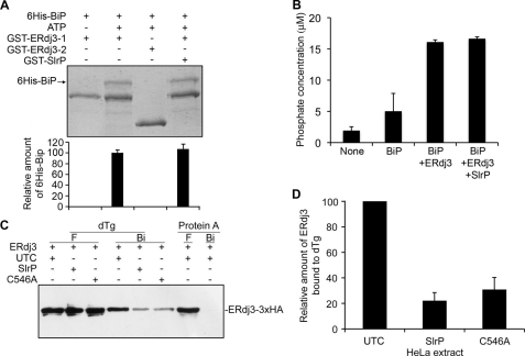
Effectors of the type III secretion systems (T3SS) are key elements in the interaction between many Gram-negative pathogens and their hosts. SlrP is an effector that is translocated into the eukaryotic host cell through the two virulence-associated T3SS of Salmonella enterica. We found previously that this effector is an E3 ubiquitin ligase for mammalian thioredoxin. Here, we identified ERdj3, an endoplasmic reticulum lumenal chaperone of the Hsp40/DnaJ family, as a new target for SlrP. Experiments with truncated forms of ERdj3 showed that domain II was essential for the interaction with SlrP. Confocal microscopy and subcellular fractionation demonstrated that, in transfected HeLa cells, SlrP was partially located in the endoplasmic reticulum. The presence of SlrP interfered with the binding of ERdj3 to a denatured substrate. Taken together, these data suggest that the role of SlrP in the interaction between Salmonella and the host cell is exerted through the modulation of the function of two independent targets: thioredoxin in the cytosol, and ERdj3 in the endoplasmic reticulum.


Figures

FIGURE 1.
FIGURE 1.
Interaction of Salmonella SlrP with human ERdj3 in the yeast two-hybrid system. A, diagram of the domains of full-length and deleted versions (1–6) of ERdj3. The domains of ERdj3 are indicated as: J, J domain; G/F, glycine-, and phenylalanine-rich domain; Ia, domain Ia; II, domain II; Ib, domain Ib; III, domain III. B, diploids were obtained by conjugation between strain AH109, containing derivatives of pGBT10 (DB-SlrP), and strain Y187, containing derivatives of pGADT7 (AD-ERdj3), as indicated. The interaction between the two hybrid proteins is shown by the growth in the absence of histidine and adenine. DB, fusion with the DNA-binding domain of Gal4; AD, fusion with the activation domain of Gal4; wt, wild-type SlrP; C546A, C546A mutant of SlrP; 1 to 6 correspond to ERdj3 subclones represented in A; −, empty vectors.
FIGURE 2.
FIGURE 2.
Interaction of ERdj3 and SlrP in vitro. A, in vitro binding of SlrP-3×FLAG to GST-ERdj3. Expression of GST and GST-ERdj3 proteins was induced with isopropyl-1-thio-β-d-galactopyranoside, and proteins were isolated from bacterial lysates by affinity chromatography with glutathione-agarose beads. 4 μg of each GST protein were incubated with lysates from 109 cfu of S. enterica serovar Typhimurium strain SV5193 (14028 slrP::3×FLAG) prepared in Nonidet P-40 lysis buffer. After washing, proteins eluted in sample buffer were resolved in 12% SDS-PAGE, blotted on nitrocellulose filters, and developed with monoclonal anti-FLAG antibody. Lysate from 108 cfu of the same strain is also included for reference. B, domain II of ERdj3 is essential for interaction with SlrP. Deleted derivatives of ERdj3 1–6 (see Fig. 1A) were expressed in fusion with GST and used for binding experiments with lysates from 109 cfu of Salmonella strain SV5193 as in A. M, molecular mass markers: visible bands correspond to 150, 100, 75, 50, 37, and 25 kDa.
FIGURE 3.
FIGURE 3.
Coimmunoprecipitation of SlrP and human ERdj3. HeLa cells were transiently co-transfected with 4 μg of two derivatives of plasmid pCS2, one expressing SlrP-3×FLAG, and the other ERdj3–3×HA (lane 1), or transfected with the plasmid expressing ERdj3–3×HA only (lane 2). Nonidet P-40 lysates from 5 × 10⁶ transfected cells were subjected to immunoprecipitation (IP, right panels) with anti-FLAG monoclonal antibodies and, after the stringent washings described under “Experimental Procedures,” resolved by 10% SDS-PAGE, transferred to nitrocellulose membranes, and developed with monoclonal anti-FLAG for the upper part of the membrane, and monoclonal anti-HA for the lower part. Lysates from 5 × 10⁵ transfected cells are shown in the left panels.
FIGURE 4.
FIGURE 4.
Colocalization of SlrP and ERdj3 in HeLa cells. A, microscopic analysis. HeLa cells transiently transfected with plasmids expressing SlrP-3×FLAG and ERdj3–3×HA were observed in a laser-scanning confocal microscope after permeabilization and staining with Alexa Fluor® 488 labeled anti-HA (green, upper panel) and Cy3-conjugated anti-FLAG (red, center panel). Superposition of the two labelings is shown in the lower panel. Yellowish staining indicates colocalization of SlrP and ERdj3 in the endoplasmic reticulum. Scale bar, 10 μm. B, detection of SlrP in the endoplasmic reticulum of HeLa-transfected cells. Total lysate (left panels) and microsomal fraction (right panels) from 107 HeLa cells stably expressing SlrP-3×FLAG were submitted to 15% SDS-PAGE, transferred to nitrocellulose membrane, and then probed with anti-FLAG monoclonal antibody to detect SlrP. The filter was also incubated with anti-ERdj3 and anti-Trx polyclonal antibodies to detect endogenous ERdj3, and endogenous Trx (a cytosolic and partially nuclear protein), respectively, as a control of the purity of the microsomal fraction. C, comparison of the migration of SlrP present in cytosolic and microsomal fractions of transfected HeLa cells. A cytosolic fraction (106 cells) and a microsomal fraction (107 cells) obtained from HeLa cells stably expressing SlrP-3×FLAG were submitted to 10% SDS-PAGE, transferred to nitrocellulose membrane and then probed with anti-FLAG monoclonal antibody to detect SlrP.
FIGURE 5.
FIGURE 5.
Effect of SlrP on ERdj3 function. A, binding of ERdj3 to BiP. 50 μg of GST-ERdj3–1 (amino acids 23–358) or GST-ERdj3–3 (amino acids 129–358) immobilized in glutathione-agarose beads were incubated with 50 μg of His6-BiP in the presence or absence of 1 mm ATP and 50 μg of GST-SlrP as indicated. After incubating at 4 °C for 2 h, the beads were washed several times with binding buffer, and bound proteins were released by boiling for 5 min in sample buffer and subjected to 10% SDS-PAGE. Proteins were detected by Coomassie Blue staining. One stained gel is shown together with a graphical representation (below) of means ± S.D., from two independent experiments, of the quantification of bands of His6-Bip. B, stimulation of BiP ATPase activity by ERdj3. ATPase assays were performed with either no additions (None), 1 μg of His6-BiP plus 2.5 μg of GST (BiP), 1 μg of His6-BiP plus 2.5 μg of GST-ERdj3–1 plus 2.5 μg of GST (BiP+ERdj3), or 1 μg of His6-BiP plus 2.5 μg of GST-ERdj3–1 plus 2.5 μg GST-SlrP (BiP+ERdj3+SlrP). The amount of released phosphate was determined after incubation at 25 °C for 75 min with a non-radioactive procedure (see “Experimental Procedures”). Values are the mean of three separate experiments with an error bar representing S.D. C, binding of ERdj3 to dTg. Lysates from HeLa cells transiently expressing ERdj3–3×HA were mixed with lysates from untransfected cells (UTC) or with lysates transfected with SlrP or SlrPCys546Ala (C546A), as indicated, and then incubated with dTg or native protein A immobilized on agarose beads. After several washes with Nonidet P-40 buffer, proteins bound to the beads (Bi) were released by boiling in sample buffer, separated by using 10% SDS-PAGE and immunoblotted with anti-HA monoclonal antibodies. Part of the initial flow-through (F) obtained after the incubations was also included. D, image quantification of the relative amount of ERdj3 bound to immobilized dTg. The results are the mean ± S.D. of three independent experiments. The highest level was set to 100 for each experiment.

Similar articles

Cited by

References

    1. Haraga A., Ohlson M. B., Miller S. I. (2008) Nat. Rev. Microbiol. 6, 53–66 - PubMed
    1. McGhie E. J., Brawn L. C., Hume P. J., Humphreys D., Koronakis V. (2009) Curr. Opin. Microbiol. 12, 117–124 - PMC - PubMed
    1. Tsolis R. M., Townsend S. M., Miao E. A., Miller S. I., Ficht T. A., Adams L. G., Bäumler A. J. (1999) Infect. Immun 67, 6385–6393 - PMC - PubMed
    1. Miao E. A., Miller S. I. (2000) Proc. Natl. Acad. Sci. U.S.A. 97, 7539–7544 - PMC - PubMed
    1. Miao E. A., Scherer C. A., Tsolis R. M., Kingsley R. A., Adams L. G., Bäumler A. J., Miller S. I. (1999) Mol. Microbiol. 34, 850–864 - PubMed

Publication types

MeSH terms

LinkOut - more resources