. 2010 May 21;285(21):16051-65.
doi: 10.1074/jbc.M109.094839. Epub 2010 Mar 24.

Structural and biochemical characterization of peroxiredoxin Qbeta from Xylella fastidiosa: catalytic mechanism and high reactivity

Affiliations

Structural and biochemical characterization of peroxiredoxin Qbeta from Xylella fastidiosa: catalytic mechanism and high reactivity

Bruno Brasil Horta et al. J Biol Chem. .

Abstract

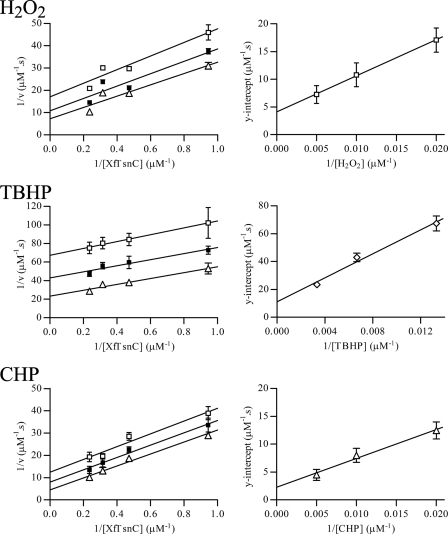
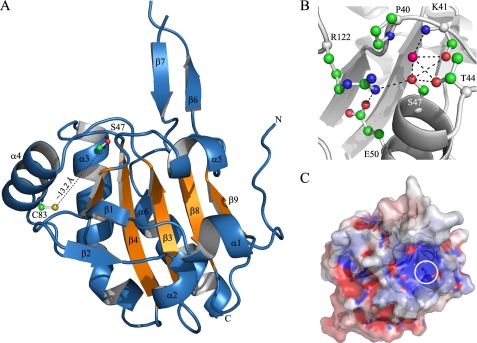
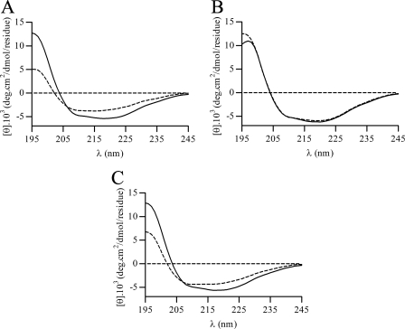
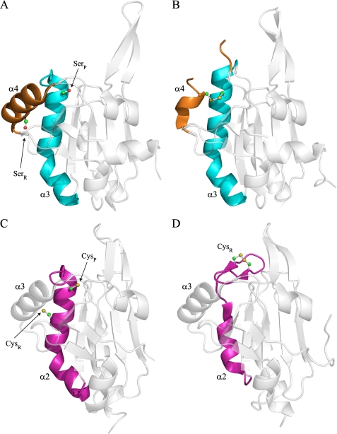
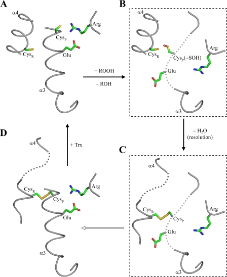
The phytopathogenic bacterium Xylella fastidiosa is the etiological agent of various plant diseases. To survive under oxidative stress imposed by the host, microorganisms express antioxidant proteins, including cysteine-based peroxidases named peroxiredoxins. This work is a comprehensive analysis of the catalysis performed by PrxQ from X. fastidiosa (XfPrxQ) that belongs to a peroxiredoxin class still poorly characterized and previously considered as moderately reactive toward hydroperoxides. Contrary to these assumptions, our competitive kinetics studies have shown that the second-order rate constants of the peroxidase reactions of XfPrxQ with hydrogen peroxide and peroxynitrite are in the order of 10(7) and 10(6) M(-1) S(-1), respectively, which are as fast as the most efficient peroxidases. The XfPrxQ disulfides were only slightly reducible by dithiothreitol; therefore, the identification of a thioredoxin system as the probable biological reductant of XfPrxQ was a relevant finding. We also showed by site-specific mutagenesis and mass spectrometry that an intramolecular disulfide bond between Cys-47 and Cys-83 is generated during the catalytic cycle. Furthermore, we elucidated the crystal structure of XfPrxQ C47S in which Ser-47 and Cys-83 lie approximately 12.3 A apart. Therefore, significant conformational changes are required for disulfide bond formation. In fact, circular dichroism data indicated that there was a significant redox-dependent unfolding of alpha-helices, which is probably triggered by the peroxidatic cysteine oxidation. Finally, we proposed a model that takes data from this work as well data as from the literature into account.


Figures

FIGURE 1.
FIGURE 1.
Peroxidase activity of XfPrxQ. A, removal of CHP was determined by DTT-dependent peroxidase assay in the presence of 5 μm XfPrxQ (closed squares), 10 μm XfTsnC (open triangles), and 5 μm XfPrxQ plus 10 μm XfTsnC (open squares). Peroxidase reactions were carried out in a reaction mixture containing 200 μm CHP and 0.5 mm DTT, at 37 °C. B, removal of hydrogen peroxide (open squares), TBHP (open triangles), and CHP (closed triangles) by XfPrxQ in DTT-dependent peroxidase assays. Peroxidase reactions were carried out in a reaction mixture containing 200 μm hydroperoxide, 0.5 mm DTT, 5 μm XfPrxQ, and 7 μm XfTsnC at 37 °C. At the indicated times, the remaining hydroperoxides were quantified in triplicate, and results are the mean ± S.D. (error bars). C, NADPH oxidation coupled to hydrogen peroxide reduction in the presence of 1 μm XfPrxQ (closed squares); 0.8 μm XfTsnC and 0.3 μm XfTrxR (open triangles); 1 μm XfPrxQ, 0.8 μm XfTsnC, and 0.3 μm XfTrxR (closed triangles); and 1 μm XfAhpC, 0.8 μm XfTsnC, and 0.3 μm XfTrxR (open squares). NADPH oxidation was measured at 340 nm in a reaction mixture containing 250 μm NADPH and 500 μm hydrogen peroxide at 37 °C. D, wild-type XfPrxQ was subjected to nonreducing SDS-PAGE under reducing conditions (lane 2), treated with 1 eq of hydrogen peroxide (lane 3) and treated with 5 eq of hydrogen peroxide (lane 4). Lane 1 shows molecular mass standards (BenchMark Protein Ladder, Invitrogen). Reduced XfPrxQ protein was treated with hydrogen peroxide at room temperature for 30 min. Approximately 4 μg of protein was loaded by lane.
FIGURE 2.
FIGURE 2.
Analysis of the primary structures of PrxQ proteins. Amino acid sequence alignments of various PrxQ from PrxQα and PrxQβ subfamilies. The red line separates PrxQ proteins into PrxQα (upper section) and Prxβ (lower section). Conserved peroxidatic and resolving cysteine residues are highlighted in blue. Conserved residues of the active site are highlighted in orange. XfPrxQ Cys-23 and Cys-101 are highlighted in green and magenta, respectively. PrxQα subfamily is represented by proteins as follows: ScDot5, S. cerevisiae DOT5 (GI: 731778); AtPrxQ, A. thaliana PrxQ (GI: 9279611); PoPrxQ, Poplar PrxQ (Populus balsamifera subsp. trichocarpa × Populus deltoides; GI: 42795441); SlPrxQ, Sedum lineare PrxQ (GI: 75336180); CdBcp, Clostridium difficile Bcp (GI: 126699430); EcBcp, E. coli Bcp (GI: 1788825); HiBcp, Haemophilus influenzae Bcp (GI: 1573220); KpBcp, K. pneumoniae Bcp (GI: 152971345); MtBcp, Mycobacterium tuberculosis Bcp (GI: 2791423); and StBcp, S. typhimurium Bcp (GI: 16765811). PrxQβ subfamily is represented by proteins as follows: RsPrxQ, Rhodobacter sphaeroides PrxQ (GI: 77387949); XcBcp, X. campestris Bcp (GI: 66768804); AvBcp, Anabaena variabilis Bcp (GI: 75906708); SyBcp, Synechocystis sp. (GI: 16329318); TeBcp, Thermosynechococcus elongatus Bcp (GI: 22298737); and XfPrxQ, Xylella fastidiosa PrxQ (GI: 9105889). The secondary structure of XfPrxQ C47S, obtained by PROCHECK (52), is shown below its sequence (green arrows representing β-sheets and red rectangles for α-helices).
FIGURE 3.
FIGURE 3.
Effects of the Cys → Ser mutations on XfPrxQ disulfide bond formation and peroxidase activity. A, removal of CHP in DTT-dependent peroxidase assay by XfPrxQ proteins as follows: wild-type (gray squares), XfPrxQ C23S (black squares), XfPrxQ C47S (gray triangles), XfPrxQ C83S (black triangles), and XfPrxQ C101S (open triangles). Peroxidase reactions were carried out in a reaction mixture containing 200 μm CHP, 2 mm DTT, and 25 μm XfPrxQ proteins at 37 °C. As a negative control, a peroxidase reaction was performed without XfPrxQ protein (open squares). At the indicated times, the remaining CHP was quantified in triplicate, and results are the mean ± S.D. (error bars). B, NADPH oxidation coupled to CHP reduction in the presence of XfPrxQ proteins as follows: wild-type (gray squares), XfPrxQ C23S (black squares), XfPrxQ C47S (gray triangles), XfPrxQ C83S (black triangles), and XfPrxQ C101S (open triangles). Assays were carried out in a reaction mixture containing 250 μm CHP, 200 μm NADPH, 1 μm XfPrxQ proteins, 1 μm E. coli Trx, and 0.1 μm E. coli TrxR at 37 °C. As a negative control, peroxidase reactions were performed without XfPrxQ protein (open squares). C, dimedone inactivation of XfPrxQ wild-type and C83S proteins. Prior to the DTT-dependent peroxidase assay, XfPrxQ proteins were incubated with 1.1 eq of hydrogen peroxide in the absence (control, black bars) or presence of 1,000 eq of dimedone (white bars) at room temperature for 30 min. Peroxidase reactions were carried out in a mixture containing 300 μm TBHP, 1 mm DTT, 1 μm E. coli Trx, and 10 μm XfPrxQ proteins at 37 °C. After 10 min, the remaining TBHP was quantified in triplicate, and results are the mean ± S.D. (error bars). Peroxidase activity was considered 100% when wild-type XfPrxQ was assayed in the absence of dimedone.
FIGURE 4.
FIGURE 4.
Localization of peroxidatic and resolving cysteines of XfPrxQ by HPLC-ESI-MS. Disulfide-containing XfPrxQ was submitted to NEM alkylation and tryptic digestion prior to HPLC-ESI-MS analyses, as described under “Experimental Procedures.” A, C-18 HPLC c of the NEM-alkylated disulfide-containing peptide mixture. The arrow indicates the retention time of the peptide scanned in C (15.66 min). B, chromatogram of the TCEP-treated mixture. The arrow indicates the retention time of the peptide scanned in D (16.97 min). Peptide elution was monitored at 214 nm. C, ion scan at 15.66 min retention time of the chromatogram showed in A. Two protonated parent ions could be used to assign a peptide formed by a disulfide bond between Cys-47 and Cys-83 (4353.07 atomic mass units). D, ion scan at 16.97 min retention time of the chromatogram showed in B. Three protonated parent ions could be used to assign a peptide containing the Cys-47 residue (3461.71 atomic mass units).
FIGURE 5.
FIGURE 5.
Steady-state bisubstrate kinetics analyses of XfPrxQ. NADPH oxidation was measured at 340 nm at 37 °C in a reaction mixture containing 0.2 mm NADPH, 5.0 μm XfTrxR, 0.2 μm XfPrxQ, various concentrations of XfTsnC (1–4 μm), and various concentrations of hydroperoxide. The data were fitted using GraphPad Prism 4. On the left side, primary plot of 1/(initial rate) versus 1/(XfTsnC concentration) at various concentrations of hydroperoxide is shown. Concentrations of hydrogen peroxide and CHP are as follows: 50 μm (open squares), 100 μm (closed squares), and 200 μm (open triangles). Concentrations of TBHP are as follows: 75 μm (open squares), 150 μm (closed squares), and 300 μm (open triangles). Each hydroperoxide concentration was analyzed in triplicate, and initial rates are the means ± S.D. (error bars). On the right side, replot of the y-intercept of primary plot versus 1/(hydroperoxide concentration).
FIGURE 6.
FIGURE 6.
Rate constant determinations of the reactions between XfPrxQ and either hydrogen peroxide or peroxynitrite by kinetic competitive approach with HRP. The reaction mixtures containing 8 μm HRP and reduced XfPrxQ in the specified concentrations were incubated with 8 μm hydrogen peroxide or peroxynitrite. After a 2-min incubation at 37 °C, formation of HRP compound I was measured at 403 nm. Each XfPrxQ protein concentration was analyzed in triplicate, and values are the means ± S.D. (error bars). A, rate constant of the reaction between wild-type (WT) XfPrxQ and hydrogen peroxide. Slope = kWT XfPrxQ = (4.53 ± 0.39) × 107 m−1 s−1. B, rate constant of the reaction between XfPrxQ C83S and hydrogen peroxide. Slope = kXfPrxQ C83S = (1.06 ± 0.07) × 107 m−1 s−1. C, variation of the second-order rate constant of the reaction between wild-type XfPrxQ and hydrogen peroxide as a function of the pH. D, Rate constant of the reaction between wild-type XfPrxQ and peroxynitrite. Slope = kWT XfPrxQ = (1.04 ± 0.03) × 106 m−1 s−1.
FIGURE 7.
FIGURE 7.
Structural features of the XfPrxQ C47S protein. A, schematic representation of the XfPrxQ C47S structure. The five-stranded mixed β-sheet of the Trx fold is colored in orange. Some elements of secondary structure are labeled, and the N and C termini are indicated. Side chains of Ser-47 and Cys-83 are shown in ball and stick mode. B, interaction network in the active site of XfPrxQ C47S. Residues are represented as balls and sticks, and atoms are colored following the CPK color scheme (carbon, green; oxygen, red; nitrogen, blue; and sulfur, yellow). The magenta ball represents water-290. Hydrogen bonds and salt bridges are indicated by dashed lines. C, XfPrxQ C47S mapped by electrostatic surface potentials (red, negatively charged; blue, positively charged), as calculated with the APBS program (65). The white circle demarcates the surface in the vicinity of the active site.
FIGURE 8.
FIGURE 8.
Conformational analysis of XfPrxQ. CD spectra in the reduced form (solid line) and treated with 1.2 eq of hydrogen peroxide (dashed line) of wild-type XfPrxQ (A), XfPrxQ C47S (B), and XfPrxQ C83S (C) proteins. Spectra were recorded at 20 °C using a protein concentration of 10 μm in 20 mm sodium phosphate buffer (pH 7.4).
FIGURE 9.
FIGURE 9.
Conformational changes in Bcp from X. campestris and A. pernix K1. A, schematic representation of Bcp C48S C84S from X. campestris (PDB code 3GKM) (54). Helix α3 and α4 that suffer conformational changes are colored in cyan and orange, respectively. Peroxidatic and resolving cysteines are substituted by serines and are indicated as SerP and SerR, respectively. The root mean square deviation for the superposition of 139 corresponding Cα atoms between XfPrxQ C47S and reduced form of Bcp from X. campestris is 0.873 Å. B, schematic representation of Bcp from X. campestris in the intramolecular disulfide bond form (PDB code 3GKK) (54). C, schematic representation of Bcp from A. pernix K1 in the reduced form (PDB code 2CX4). Helix α2 that suffer conformational change is colored in magenta. Peroxidatic and resolving cysteines are indicated as CysP and CysR, respectively. The root mean square deviation for the superposition of 139 corresponding Cα atoms between XfPrxQ C47S and Bcp from A. pernix K1 is 1.785 Å. D, schematic representation of the oxidized form of Bcp from A. pernix K1 (PDB code 2CX3). Residues involved in intramolecular disulfide bond are represented as balls and sticks, and atoms are colored following CPK color scheme.
FIGURE 10.
FIGURE 10.
Structurally detailed model of conformational changes in the PrxQβ catalytic cycle. Proposed sequence of structure snapshots along the catalytic cycle of PrxQ subfamily β-proteins. Each panel represents a different species of the proposed model. A, reduced species based on the determined crystal structure of XfPrxQ C47S. B and C, these species in dashed boxes represent hypothetical conformational intermediates based on CD data presented here. D, oxidized species based on the determined crystal structure of Bcp from X. campestris (PDB code 3GKK) (54). Peroxidatic and resolving cysteines are indicated as CysP and CysR, respectively. Residues are represented as sticks, and atoms are colored following the CPK color scheme.

Similar articles

Cited by

References

    1. Simpson A. J., Reinach F. C., Arruda P., Abreu F. A., Acencio M., Alvarenga R., Alves L. M., Araya J. E., Baia G. S., Baptista C. S., Barros M. H., Bonaccorsi E. D., Bordin S., Bové J. M., Briones M. R., Bueno M. R., Camargo A. A., Camargo L. E., Carraro D. M., Carrer H., Colauto N. B., Colombo C., Costa F. F., Costa M. C., Costa-Neto C. M., Coutinho L. L., Cristofani M., Dias-Neto E., Docena C., El-Dorry H., Facincani A. P., Ferreira A. J., Ferreira V. C., Ferro J. A., Fraga J. S., França S. C., Franco M. C., Frohme M., Furlan L. R., Garnier M., Goldman G. H., Goldman M. H., Gomes S. L., Gruber A., Ho P. L., Hoheisel J. D., Junqueira M. L., Kemper E. L., Kitajima J. P., Krieger J. E., Kuramae E. E., Laigret F., Lambais M. R., Leite L. C., Lemos E. G., Lemos M. V., Lopes S. A., Lopes C. R., Machado J. A., Machado M. A., Madeira A. M., Madeira H. M., Marino C. L., Marques M. V., Martins E. A., Martins E. M., Matsukuma A. Y., Menck C. F., Miracca E. C., Miyaki C. Y., Monteriro-Vitorello C. B., Moon D. H., Nagai M. A., Nascimento A. L., Netto L. E., Nhani A., Jr., Nobrega F. G., Nunes L. R., Oliveira M. A., de Oliveira M. C., de Oliveira R. C., Palmieri D. A., Paris A., Peixoto B. R., Pereira G. A., Pereira H. A., Jr., Pesquero J. B., Quaggio R. B., Roberto P. G., Rodrigues V., de M, Rosa A. J., de Rosa V. E., Jr., de Sá R. G., Santelli R. V., Sawasaki H. E., da Silva A. C., da Silva A. M., da Silva F. R., da Silva W. A., Jr., da Silveira J. F., Silvestri M. L., Siqueira W. J., de Souza A. A., de Souza A. P., Terenzi M. F., Truffi D., Tsai S. M., Tsuhako M. H., Vallada H., Van Sluys M. A., Verjovski-Almeida S., Vettore A. L., Zago M. A., Zatz M., Meidanis J., Setubal J. C. (2000) Nature 406, 151–159 - PubMed
    1. Amaro A. A., Maia M. L., Gonzales M. A. (1998) in Citrus Variegated Chlorosis (Donadio L. C., Moreira C. S. eds) pp. 123–139, Estação Experimental de Citricultura, Bebedouro, SP, Brazil
    1. Hendson M., Purcell A. H., Chen D., Smart C., Guilhabert M., Kirkpatrick B. (2001) Appl. Environ. Microbiol. 67, 895–903 - PMC - PubMed
    1. Wrzaczek M., Brosché M., Kollist H., Kangasjärvi J. (2009) Proc. Natl. Acad. Sci. U.S.A. 106, 5412–5417 - PMC - PubMed
    1. Tenhaken R., Levine A., Brisson L. F., Dixon R. A., Lamb C. (1995) Proc. Natl. Acad. Sci. U.S.A. 92, 4158–4163 - PMC - PubMed

Publication types

LinkOut - more resources