. 2010 May 28;285(22):16931-41.
doi: 10.1074/jbc.M109.099036. Epub 2010 Mar 24.

A novel cross-talk in diacylglycerol signaling: the Rac-GAP beta2-chimaerin is negatively regulated by protein kinase Cdelta-mediated phosphorylation

Affiliations

A novel cross-talk in diacylglycerol signaling: the Rac-GAP beta2-chimaerin is negatively regulated by protein kinase Cdelta-mediated phosphorylation

Erin M Griner et al. J Biol Chem. .

Abstract

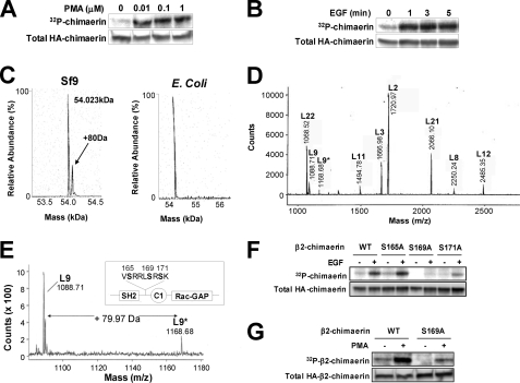
Although the family of chimaerin Rac-GAPs has recently gained significant attention for their involvement in development, cancer, and neuritogenesis, little is known about their molecular regulation. Chimaerins are activated by the lipid second messenger diacylglycerol via their C1 domain upon activation of tyrosine kinase receptors, thereby restricting the magnitude of Rac signaling in a receptor-regulated manner. Here we identified a novel regulatory mechanism for beta2-chimaerin via phosphorylation. Epidermal growth factor or the phorbol ester phorbol 12-myristate 13-acetate caused rapid phosphorylation of beta2-chimaerin on Ser(169) located in the SH2-C1 domain linker region via protein kinase Cdelta, which retained beta2-chimaerin in the cytosol and prevented its C1 domain-mediated translocation to membranes. Furthermore, despite the fact that Ser(169) phosphorylation did not alter intrinsic Rac-GAP activity in vitro, a non-phosphorylatable beta2-chimaerin mutant was highly sensitive to translocation, and displayed enhanced association with activated Rac, enhanced Rac-GAP activity, and anti-migratory properties when expressed in cells. Our results not only revealed a novel regulatory mechanism that facilitates Rac activation, but also identified a novel mechanism of cross-talk between diacylglycerol receptors that restricts beta2-chimaerin relocalization and activation.


Figures

FIGURE 1.
FIGURE 1.
β2-Chimaerin is phosphorylated on Ser169 in response to EGF or PMA. A and B, serum-starved COS-1 cells labeled with 32Pi were treated with PMA (10 min) (A) or EGF (100 ng/ml) (B). Phosphorylation was monitored by autoradiography, and HA-β2-chimaerin expression was assessed by Western blotting. C, recombinant β2-chimaerin purified from Sf9 cells (left) or E. coli (right) was subjected to mass spectroscopy. D, spectrum obtained upon MALDI-TOF mass spectroscopy analysis of Lys-C-digested β2-chimaerin isolated from Sf9 cells. E, mass spectrum of putative phosphopeptide. Inset, localization of the putative phosphorylation sites in β2-chimaerin. F and G, COS-1 cells expressing HA-β2-chimaerin Ser to Ala mutants were labeled with 32Pi and treated with EGF (100 ng/ml, 5 min) (F) or PMA (1 μm, 10 min) (G). Phosphorylation was monitored by autoradiography.
FIGURE 2.
FIGURE 2.
Analysis of β2-chimaerin phosphorylation and stoichiometry of phosphorylation using a phosphospecific antibody. A and B, Ser169 phosphorylation of HA-β2-chimaerin was assessed in COS-1 cells using a phospho-Ser169 specific antibody following EGF (100 ng/ml) (A) or PMA (1 μm) (B) treatments. Top, representative Western blots. Bottom, data relative to t = 0 min presented as mean ± S.E. C, serum-starved COS-1 cells expressing HA-β2-chimaerin AdV were treated with EGF (100 ng/ml, 5 min). Cell lysates were subjected to two-dimensional gel electrophoresis and Western blotting. Left, representative Western blots. Right, analysis of EGF-treated samples expressed as percentage of non-phosphorylated or phosphorylated protein (relative to total protein) expressed as the mean ± S.E. (n = 5). D, primary mouse cerebellar granule neurons were treated with brain-derived neurotrophic factor (BDNF) (100 ng/ml) for the indicated times and assessed for Ser169 phosphorylation of endogenous β2-chimaerin.
FIGURE 3.
FIGURE 3.
PKCδ phosphorylates β2-chimaerin on Ser169. A, effect of GF (0–10 μm, 1 h) on EGF-stimulated phosphorylation (100 ng/ml, 5 min) of HA-β2-chimaerin expressed in COS-1 cells, as measured by autoradiography. B, expression of PKC isozymes in HeLa cells following RNA interference; NTS, non-targeting sequence. C, HeLa cells subjected to RNA interference depletion of PKCs were infected with HA-β2-chimaerin AdV, serum starved, and treated with EGF. Top, representative Western blot. Bottom, data expressed as % of NTS (+ EGF) are presented as mean ± S.E. (n = 4), *, p < 0.05. D, expression of PKC isozymes in stable HeLa cell lines created using shRNA lentiviruses directed against PKCδ or a NTS. E, serum-starved cell lines in D infected with HA-β2-chimaerin AdV were treated with EGF. Top, representative Western blot. Bottom, data expressed as % of NTS (+ EGF) presented as mean ± S.E. (n = 3), **, p < 0.01; ***, p < 0.005. F, serum-starved COS-1 cells infected with LacZ or PKCδ AdVs were stimulated with EGF. Top, representative Western blot. Bottom, data expressed relative to LacZ (− EGF) presented as mean ± S.E. (n = 4), *, p < 0.05. G, in vitro phosphorylation of Ser169 of β2-chimaerin by recombinant PKCδ was measured by Western blot. siRNA, small interfering RNA.
FIGURE 4.
FIGURE 4.
S169A-β2-chimaerin has enhanced GAP activity in cells. A and B, serum-starved HeLa cells infected with LacZ, WT, or S169A-β2-chimaerin AdVs were treated with EGF (100 ng/ml, 1 min). Rac1-GTP levels (A) or RhoA-GTP levels (B) were assessed using pulldown assays. Top, results expressed as fold-change relative to LacZ (− EGF) presented as the mean ± S.E. (n = 3). **, p < 0.01. Bottom, representative Western blots. C and D, FBS-directed migration of HeLa cells expressing LacZ, WT-, or S169A-β2-chimaerin was determined using a Boyden chamber. Cells in D also co-expressed V12Rac1 as indicated. Left, representative images of migrated cells. Right, quantification of the number of migrated cells and Western blots showing the expression of chimaerin proteins. Results are expressed as mean ± S.E. (n = 3). ***, p < 0.0001.
FIGURE 5.
FIGURE 5.
Phosphorylated β2-chimaerin is localized in the cytosol. A, lysates from COS-1 cells expressing HA-β2-chimaerin were fractionated. Phosphorylation was monitored by Western blot. B, COS-1 cells expressing WT- or P223A-β2-chimaerin were treated with GF (10 μm, 1 h), then with PMA (0–10 μm, 5 min) and fractionated. Chimaerins were detected by Western blot. Numbers below the blot represent the fold-change in protein present in the insoluble fraction with respect to WT, no PMA. C, COS-1 cells expressing the indicated β2-chimaerin mutants were treated with EGF (100 ng/ml, 5 min). Phosphorylation was monitored by Western blot. Left, representative Western blot. Right, data presented relative to WT (+ EGF) expressed as mean ± S.E.
FIGURE 6.
FIGURE 6.
Phosphorylated β2-chimaerin is unable to translocate to membranes in response to PMA. A, COS-1 cells expressing WT or mutant β2-chimaerin were treated with PMA (1 μm, 5 min) with or without GF (10 μm, 1 h). Cell lysates were fractionated and β2-chimaerin was detected by Western blot. B, GFP-tagged WT- or S169A-β2-chimaerin localization in COS-1 cells in response to PMA (1 μm, 5 min) ± GF (10 μm, 1 h) was monitored by confocal microscopy.
FIGURE 7.
FIGURE 7.
S169A-β2-chimaerin does not have enhanced Rac-GAP activity in vitro. In vitro Rac-GAP activity of recombinant chimaerin proteins is shown. Results are expressed as mean ± S.E. (n = 3).
FIGURE 8.
FIGURE 8.
Enhanced association of S169A-β2-chimaerin to V12Rac1. A and B, lysates from COS-1 cells co-expressing GST or GST-V12Rac1 and the indicated β2-chimaerin mutants were subjected to GST pulldown. HA-β2-chimaerin bound to GST beads was detected by Western blot. Left, representative Western blots. Right, data presented relative to WT- (A) or S169A-β2-chimaerin (B) expressed as mean ± S.E. (n = 4–5), **, p < 0.01; ***, p < 0.005.
FIGURE 9.
FIGURE 9.
Proposed model for β2-chimaerin regulation by Ser169 phosphorylation. Under resting conditions, β2-chimaerin remains in an inactive, closed conformation in the cytosol. Upon EGFR stimulation, DAG generated via phospholipase Cγ activates PKCδ, which in turn phosphorylates β2-chimaerin on Ser169. Phosphorylated β2-chimaerin is unable to translocate to membranes and remains inactive in the cytosol. On the other hand, non-phosphorylated β2-chimaerin is subject to allosteric activation by DAG and acidic phospholipids in the plasma membrane, where it inactivates Rac.

Similar articles

Cited by

References

    1. Blumberg P. M., Delclos K. B., Dunn J. A., Jaken S., Leach K. L., Yeh E. (1983) Ann. N.Y. Acad. Sci. 407, 303–315 - PubMed
    1. Colón-González F., Kazanietz M. G. (2006) Biochim. Biophys. Acta 1761, 827–837 - PubMed
    1. Griner E. M., Kazanietz M. G. (2007) Nat. Rev. Cancer 7, 281–294 - PubMed
    1. Newton A. C. (2003) Biochem. J. 370, 361–371 - PMC - PubMed
    1. Parekh D. B., Ziegler W., Parker P. J. (2000) EMBO J. 19, 496–503 - PMC - PubMed

Publication types

MeSH terms