

The mutation N528S in the von Willebrand factor (VWF) propeptide causes defective multimerization and storage of VWF
- PMID: 20335223
- PMCID: PMC2881501
- DOI: 10.1182/blood-2009-09-244327
The mutation N528S in the von Willebrand factor (VWF) propeptide causes defective multimerization and storage of VWF
Abstract
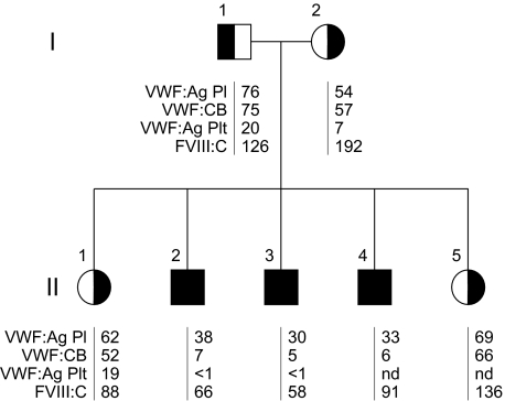
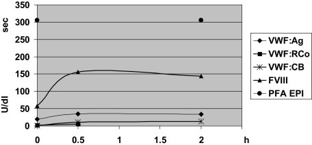
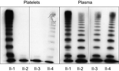
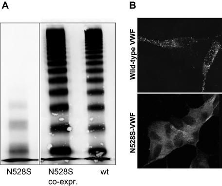
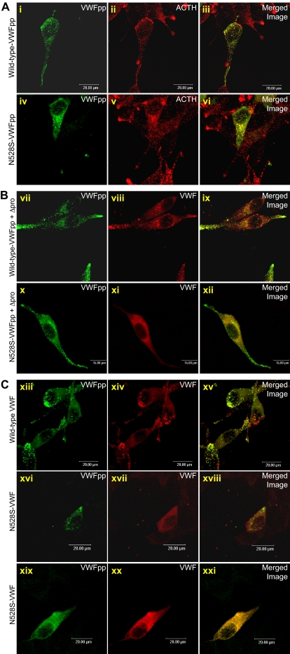
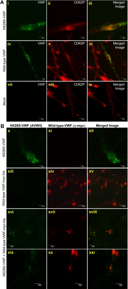
We characterized a consanguineous Turkish family suffering from von Willebrand disease (VWD) with significant mucocutaneous and joint bleeding. The relative reduction of large plasma von Willebrand factor (VWF) multimers and the absent VWF triplet structure was consistent with type 2A (phenotype IIC) VWD. Surprisingly, platelet VWF was completely deficient of multimers beyond the VWF protomer, suggesting defective alpha-granular storage of larger multimers. Patients were nearly unresponsive to desmopressin acetate, consistent with a lack of regulated VWF release from endothelial cell Weibel-Palade bodies, suggesting defective storage also in endothelial cells. We identified an N528S homozygous mutation in the VWF propeptide D2 domain, predicting the introduction of an additional N-glycosylation site at amino acid 526 in close vicinity to a "CGLC" disulphide isomerase consensus sequence. Expression studies in mammalian cells demonstrated that N528S-VWF was neither normally multimerized nor trafficked to storage granules. However, propeptide containing the N528S mutation trafficked normally to storage granules. Our data indicate that the patients' phenotype is the result of defective multimerization, storage, and secretion. In addition, we have identified a potentially novel pathogenic mechanism of VWD, namely a transportation and storage defect of mature VWF due to defective interaction with its transporter, the mutant propeptide.
Figures
Similar articles
-
Identification and characterization of the elusive mutation causing the historical von Willebrand Disease type IIC Miami.J Thromb Haemost. 2016 Sep;14(9):1725-35. doi: 10.1111/jth.13398. Epub 2016 Aug 20. J Thromb Haemost. 2016. PMID: 27344059 Free PMC article.
-
A new candidate mutation (N528S) within the von Willebrand factor propeptide identified in a Japanese patient with phenotype IIC of von Willebrand disease.Eur J Haematol. 1998 Aug;61(2):145-8. doi: 10.1111/j.1600-0609.1998.tb01076.x. Eur J Haematol. 1998. PMID: 9714529
-
Laboratory diagnosis of von Willebrand disease type 1/2E (2A subtype IIE), type 1 Vicenza and mild type 1 caused by mutations in the D3, D4, B1-B3 and C1-C2 domains of the von Willebrand factor gene. Role of von Willebrand factor multimers and the von Willebrand factor propeptide/antigen ratio.Acta Haematol. 2009;121(2-3):128-38. doi: 10.1159/000214853. Epub 2009 Jun 8. Acta Haematol. 2009. PMID: 19506359 Review.
-
Clinical and laboratory phenotype variability in type 2M von Willebrand disease.J Thromb Haemost. 2017 Aug;15(8):1559-1566. doi: 10.1111/jth.13742. Epub 2017 Jun 23. J Thromb Haemost. 2017. PMID: 28544236 Free PMC article.
-
Molecular genetics of type 2 von Willebrand disease.Int J Hematol. 2002 Jan;75(1):9-18. doi: 10.1007/BF02981973. Int J Hematol. 2002. PMID: 11843298 Review.
Cited by
-
Interpreting functional effects of coding variants: challenges in proteome-scale prediction, annotation and assessment.Brief Bioinform. 2016 Sep;17(5):841-62. doi: 10.1093/bib/bbv084. Epub 2015 Oct 22. Brief Bioinform. 2016. PMID: 26494363 Free PMC article. Review.
-
Identification and characterization of the elusive mutation causing the historical von Willebrand Disease type IIC Miami.J Thromb Haemost. 2016 Sep;14(9):1725-35. doi: 10.1111/jth.13398. Epub 2016 Aug 20. J Thromb Haemost. 2016. PMID: 27344059 Free PMC article.
-
Angiodysplasia in von Willebrand Disease: Understanding the Clinical and Basic Science.Semin Thromb Hemost. 2017 Sep;43(6):572-580. doi: 10.1055/s-0037-1599145. Epub 2017 May 5. Semin Thromb Hemost. 2017. PMID: 28476066 Free PMC article. Review.
-
Mutations in the D'D3 region of VWF traditionally associated with type 1 VWD lead to quantitative and qualitative deficiencies of VWF.Thromb Res. 2016 Sep;145:112-8. doi: 10.1016/j.thromres.2016.08.009. Epub 2016 Aug 10. Thromb Res. 2016. PMID: 27533707 Free PMC article.
-
Update on von Willebrand factor multimers: focus on high-molecular-weight multimers and their role in hemostasis.Blood Coagul Fibrinolysis. 2014 Apr;25(3):206-16. doi: 10.1097/MBC.0000000000000065. Blood Coagul Fibrinolysis. 2014. PMID: 24448155 Free PMC article. Review.
References
-
- Sadler JE, Mannucci PM, Berntorp E, et al. Impact, diagnosis and treatment of von Willebrand disease. Thromb Haemost. 2000;84(2):160–174. - PubMed
-
- Sadler JE, Budde U, Eikenboom JC, et al. Update on the pathophysiology and classification of von Willebrand disease: a report of the Subcommittee on von Willebrand Factor. J Thromb Haemost. 2006;4(10):2103–2114. - PubMed
Publication types
MeSH terms
Substances
Grants and funding
LinkOut - more resources
Full Text Sources
Molecular Biology Databases
Miscellaneous
