. 2010 Jun;84(11):5670-7.
doi: 10.1128/JVI.00272-10. Epub 2010 Mar 24.

Lambda interferon renders epithelial cells of the respiratory and gastrointestinal tracts resistant to viral infections

Affiliations

Lambda interferon renders epithelial cells of the respiratory and gastrointestinal tracts resistant to viral infections

Markus Mordstein et al. J Virol. 2010 Jun.

Abstract

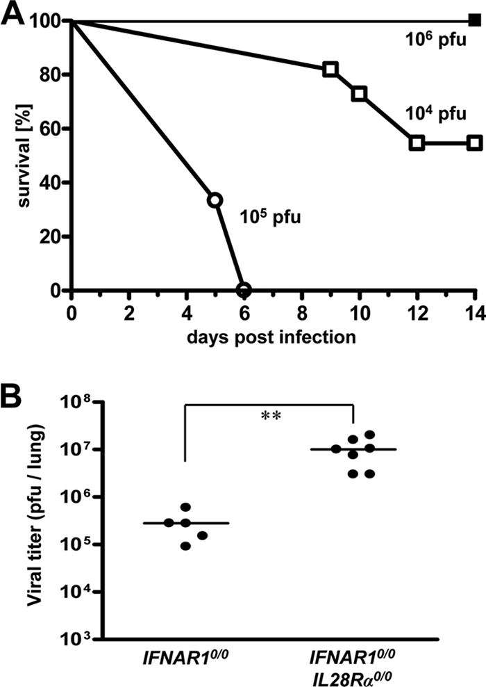
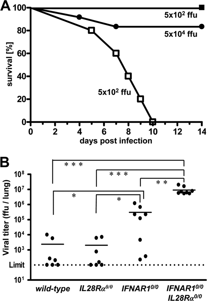
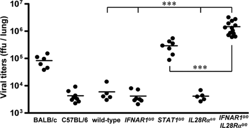
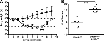
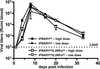
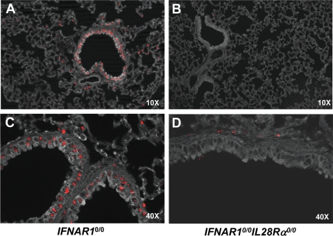
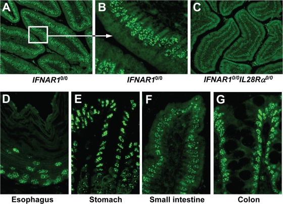
Virus-infected cells secrete a broad range of interferons (IFN) which confer resistance to yet uninfected cells by triggering the synthesis of antiviral factors. The relative contributions of the various IFN subtypes to innate immunity against virus infections remain elusive. IFN-alpha, IFN-beta, and other type I IFN molecules signal through a common, universally expressed cell surface receptor, whereas type III IFN (IFN-lambda) uses a distinct cell-type-specific receptor complex for signaling. Using mice lacking functional receptors for type I IFN, type III IFN, or both, we found that IFN-lambda plays an important role in the defense against several human pathogens that infect the respiratory tract, such as influenza A virus, influenza B virus, respiratory syncytial virus, human metapneumovirus, and severe acute respiratory syndrome (SARS) coronavirus. These viruses were more pathogenic and replicated to higher titers in the lungs of mice lacking both IFN receptors than in mice with single IFN receptor defects. In contrast, Lassa fever virus, which infects via the respiratory tract but primarily replicates in the liver, was not influenced by the IFN-lambda receptor defect. Careful analysis revealed that expression of functional IFN-lambda receptor complexes in the lung and intestinal tract is restricted to epithelial cells and a few other, undefined cell types. Interestingly, we found that SARS coronavirus was present in feces from infected mice lacking receptors for both type I and type III IFN but not in those from mice lacking single receptors, supporting the view that IFN-lambda contributes to the control of viral infections in epithelial cells of both respiratory and gastrointestinal tracts.


Figures

FIG. 1.
FIG. 1.
Mice lacking functional receptors for both IFN-α/β and IFN-λ exhibit high susceptibility toward influenza virus strain A/HH/05/2009 (H1N1). (A) Survival of IFNAR10/0 mice (filled squares; n = 4) and IFNAR10/0IL28Rα0/0 double-knockout mice (open symbols; 104 PFU [n = 11] and 105 PFU [n = 3]) after intranasal infection with the indicated doses of virus. (B) Virus titers in lungs at 48 h following intranasal infection with 103 PFU of virus. Combined data for two independent experiments are shown. Each dot represents the data for one animal. **, P < 0.01.
FIG. 2.
FIG. 2.
Mice lacking functional receptors for both IFN-α/β and IFN-λ exhibit high susceptibility toward influenza virus strain B/Lee/40. (A) Survival of IFNAR10/0 mice (filled symbols; 5 × 10² PFU [n = 7] and 5 × 10⁴ PFU [n = 10]) and IFNAR10/0IL28Rα0/0 double-knockout mice (open squares; n = 4) after intranasal infection with the indicated doses of virus. Combined data for two independent experiments are shown. (B) Virus titers in lungs at 72 h following intranasal infection with 5 × 10⁴ FFU of virus. Combined data for several independent experiments are shown. Each dot represents the data for one animal. *, P < 0.05; **, P < 0.01; ***, P < 0.001.
FIG. 3.
FIG. 3.
Mice lacking functional receptors for both IFN-α/β and IFN-λ exhibit high susceptibility toward RSV. Virus titers in lungs at day 4 following intranasal infection with 8 × 10⁶ FFU of RSV. Wild-type mice and C57BL/6 mice differ only by the presence or absence of functional Mx1 alleles. Combined data for several independent experiments are shown. Each dot represents the data for one animal. ***, P < 0.001.
FIG. 4.
FIG. 4.
Mice lacking functional receptors for both IFN-α/β and IFN-λ exhibit high susceptibility toward HMPV. (A) Body weight changes of IFNAR10/0 (n = 4) and IFNAR10/0IL28Rα0/0 (n = 5) mice intranasally infected with 105 PFU of HMPV. Crosses indicate that animals had to be killed due to severe symptoms. (B) HMPV-RNA load in lungs at day 4 following intranasal infection with 105 PFU of HMPV was determined by qRT-PCR. Combined data for two independent experiments are shown. Each dot represents the data for one animal. ***, P < 0.001.
FIG. 5.
FIG. 5.
Lassa fever virus replication is not affected by IFN-λ. IFNAR10/0 and IFNAR10/0IL28Rα0/0 mice were infected by the intranasal route with either 3 × 10⁵ PFU (high dose) or 3 × 10³ PFU (low dose) of Lassa fever virus. Blood samples were taken at the indicated times postinfection and tested for presence of virus. Each group consisted of three mice, except the IFNAR10/0IL28Rα0/0 low-dose group, which consisted of two mice.
FIG. 6.
FIG. 6.
Lung epithelial cells express functional IFN-λ receptor complexes. IFNAR10/0 (A, C) and IFNAR10/0IL28Rα0/0 (B, D) mice were intranasally infected with 5 × 10⁴ PFU of SC35M-ΔNS1, a potent inducer of type I and type III IFN (26). At 20 h postinfection, the lungs were removed and stained for Mx1 by immunohistofluorescence. (A, B) Low magnification overview. Mx1-positive cells (stained nuclei) are mainly clustered around bronchioles in IFNAR10/0 mice but mostly absent in IFNAR10/0IL28Rα0/0 mice. (B, D) High magnification of bronchioles and surrounding tissue. Epithelial cells were prominently stained for Mx1 in IFNAR10/0 but not in IFNAR10/0IL28Rα0/0 mice.
FIG. 7.
FIG. 7.
Epithelial cells of the intestine express functional IFN-λ receptor complexes. The response to in vivo electroporation of a plasmid encoding mouse IFN-λ3 was monitored by immunohistofluorescence staining for Mx1. The duodenum (section of the villi) of IFNAR10/0 mice is shown in low (A) and high (B) magnification. Prominent labeling of nuclear Mx1 is detected mainly in epithelial cells of the villi. (C) No such staining was observed in IFNAR10/0IL28Rα0/0 mice, which cannot respond to IFN-λ. Strongly stained epithelial cells were detected in all regions of the gastrointestinal tract of IFN-λ-treated IFNAR10/0 mice, including esophagus (D), stomach (E), small intestine (F), and colon (G).
FIG. 8.
FIG. 8.
IFN-λ contributes to restriction of SARS-CoV replication in both lungs and intestinal tracts of mice. (A) Virus titers in lungs of various mouse lines at day 4 following intranasal infection with 106 PFU of SARS-CoV per animal. Combined data for two independent experiments are shown. Each dot represents the data for one animal. *, P < 0.05; ***, P < 0.001. (B) At days 4 and 14 postinfection, RNA samples extracted from 20 droppings of cages harboring the different mouse lines were analyzed by qRT-PCR for SARS-CoV. (C) RNA samples from intestines of infected wild-type and IFNAR10/0IL28Rα0/0 double-knockout mice were analyzed by qRT-PCR for SARS-CoV. Viral RNA levels were calculated using appropriate standards.

References

    1. Ank, N., M. B. Iversen, C. Bartholdy, P. Staeheli, R. Hartmann, U. B. Jensen, F. Dagnaes-Hansen, A. R. Thomsen, Z. Chen, H. Haugen, K. Klucher, and S. R. Paludan. 2008. An important role for type III interferon (IFN-lambda/IL-28) in TLR-induced antiviral activity. J. Immunol. 180:2474-2485. - PubMed
    1. Asper, M., T. Sternsdorf, M. Hass, C. Drosten, A. Rhode, H. Schmitz, and S. Gunther. 2004. Inhibition of different Lassa virus strains by alpha and gamma interferons and comparison with a less pathogenic arenavirus. J. Virol. 78:3162-3169. - PMC - PubMed
    1. Bartlett, N. W., K. Buttigieg, S. V. Kotenko, and G. L. Smith. 2005. Murine interferon lambdas (type III interferons) exhibit potent antiviral activity in vivo in a poxvirus infection model. J. Gen. Virol. 86:1589-1596. - PubMed
    1. Bouloy, M., C. Janzen, P. Vialat, H. Khun, J. Pavlovic, M. Huerre, and O. Haller. 2001. Genetic evidence for an interferon-antagonistic function of rift valley fever virus nonstructural protein NSs. J. Virol. 75:1371-1377. - PMC - PubMed
    1. Cervantes-Barragan, L., R. Zust, F. Weber, M. Spiegel, K. S. Lang, S. Akira, V. Thiel, and B. Ludewig. 2007. Control of coronavirus infection through plasmacytoid dendritic-cell-derived type I interferon. Blood 109:1131-1137. - PMC - PubMed

Publication types