. 2010 Jul;84(13):6782-98.
doi: 10.1128/JVI.01757-09. Epub 2010 Mar 24.

Rotaviruses associate with cellular lipid droplet components to replicate in viroplasms, and compounds disrupting or blocking lipid droplets inhibit viroplasm formation and viral replication

Affiliations

Rotaviruses associate with cellular lipid droplet components to replicate in viroplasms, and compounds disrupting or blocking lipid droplets inhibit viroplasm formation and viral replication

Winsome Cheung et al. J Virol. 2010 Jul.

Abstract

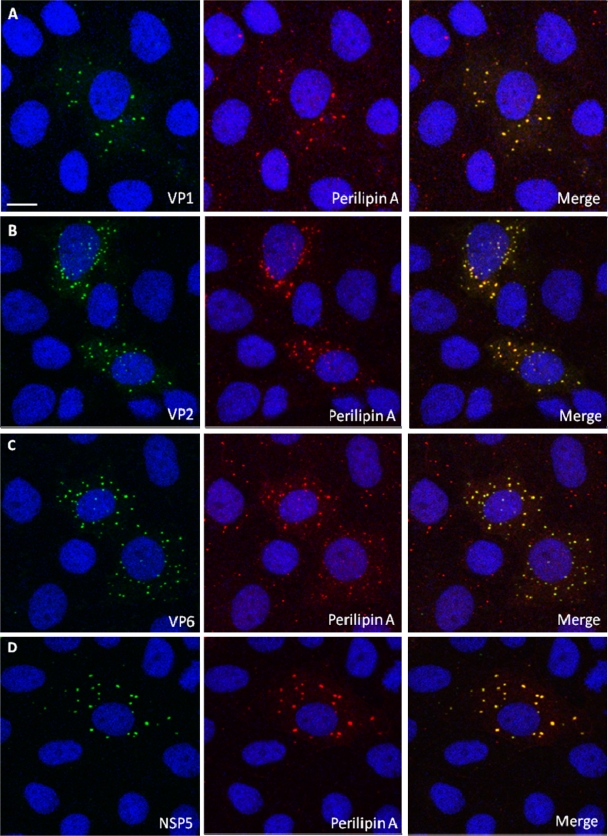
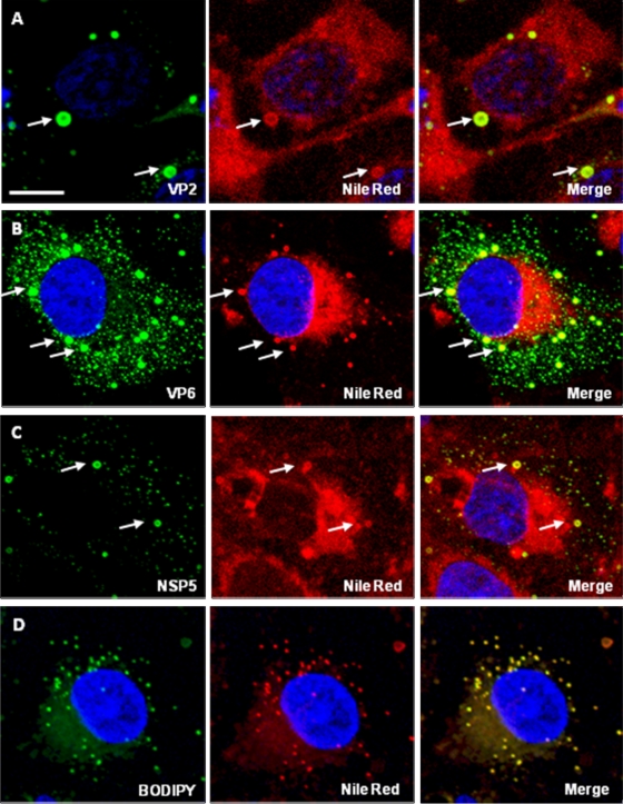
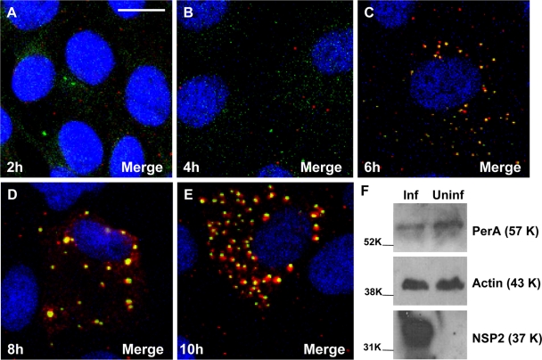
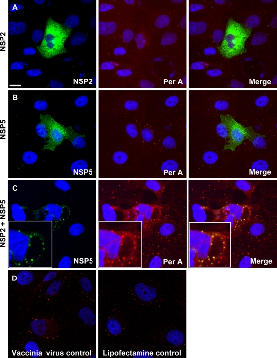
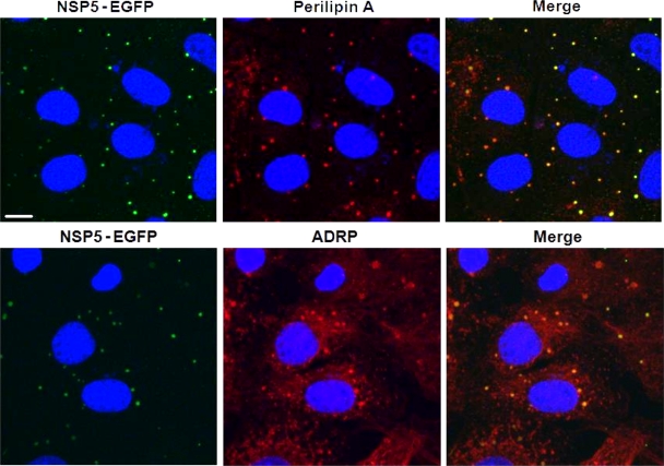
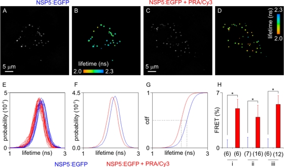
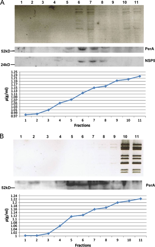
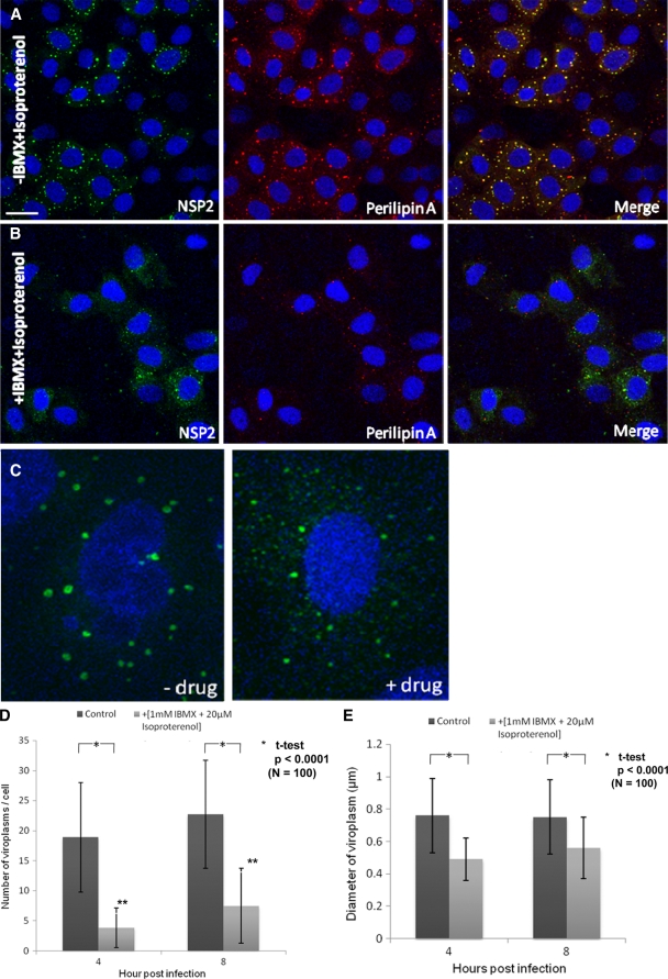
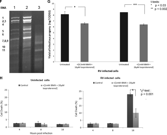
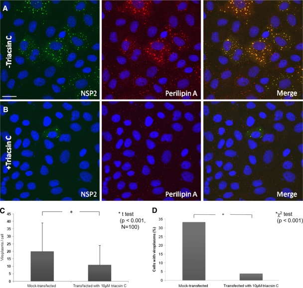
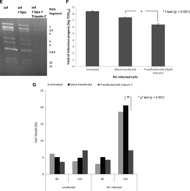
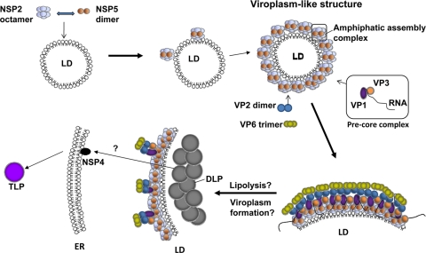
Rotaviruses are a major cause of acute gastroenteritis in children worldwide. Early stages of rotavirus assembly in infected cells occur in viroplasms. Confocal microscopy demonstrated that viroplasms associate with lipids and proteins (perilipin A, ADRP) characteristic of lipid droplets (LDs). LD-associated proteins were also found to colocalize with viroplasms containing a rotaviral NSP5-enhanced green fluorescent protein (EGFP) fusion protein and with viroplasm-like structures in uninfected cells coexpressing viral NSP2 and NSP5. Close spatial proximity of NSP5-EGFP and cellular perilipin A was confirmed by fluorescence resonance energy transfer. Viroplasms appear to recruit LD components during the time course of rotavirus infection. NSP5-specific siRNA blocked association of perilipin A with NSP5 in viroplasms. Viral double-stranded RNA (dsRNA), NSP5, and perilipin A cosedimented in low-density gradient fractions of rotavirus-infected cell extracts. Chemical compounds interfering with LD formation (isoproterenol plus isobutylmethylxanthine; triacsin C) decreased the number of viroplasms and inhibited dsRNA replication and the production of infectious progeny virus; this effect correlated with significant protection of cells from virus-associated cytopathicity. Rotaviruses represent a genus of another virus family utilizing LD components for replication, pointing at novel therapeutic targets for these pathogens.


Figures

FIG. 1.
FIG. 1.
NSP2 colocalizes with LD-associated proteins perilipin A and ADRP in viroplasms. Confocal images of rotavirus-infected MA104 cells at 8 h p.i. were obtained. (A and B) Viroplasms were detected with anti-NSP2 antibodies, followed by visualization with Alexa Fluor 488 (green)-labeled secondary antibody, whereas LD-associated proteins were detected with anti-perilipin A (A) and anti-ADRP antibodies (B), followed by reaction with Alexa Fluor 633 (red)-labeled secondary antibody. An individual viroplasm of panel A has been magnified and is shown in the inserts. (C) Confocal images of uninfected MA104 cells stained for perilipin A or ADRP and both for NSP2 according to the procedure used in panels A and B. Uninfected cells only show LDs. Scale bar, 10 μm.
FIG. 2.
FIG. 2.
Perilipin A colocalizes with VP1, VP2, VP6, and NSP5 in viroplasms. Shown are CM images of rotavirus-infected MA104 cells at 6 h p.i. Viroplasms were detected with anti-perilipin A antibodies visualized by Alexa Fluor 633 (red)-conjugated secondary antibodies, while VP1 (A), VP2 (B), VP6 (C), and NSP5 (D) were detected by monospecific antibodies raised in guinea pigs, followed by visualization with secondary antibodies conjugated with Alexa-Fluor 488 (green). Scale bar, 10 μm.
FIG. 3.
FIG. 3.
Rotavirus proteins colocalize with lipids in viroplasms. Confocal images of rotavirus-infected Cos-7 cells costained with Nile Red and antibodies to VP2 (A, 8 h p.i.), VP6 (B, 8 h p.i.), and NSP5 (C, 4 h p.i.). (D) uninfected cell controls stained with Bodipy (left) and Nile Red (center). The merged picture is shown on the right. Scale bar, 10 μm.
FIG. 4.
FIG. 4.
(A to E) Time course analysis of viroplasm development in rotavirus-infected MA104 cells showing the recruitment of LD-associated proteins. MA104 cells on coverslips were infected with rotavirus RF strain at an MOI of 1, and cells were fixed at 2, 4, 6, 8, and 10 h p.i. as indicated. Viroplasms were detected with anti-perilipin A antibodies visualized by Alexa Fluor 633 (red)-conjugated secondary antibodies, while NSP5 was detected by monospecific antibodies raised in guinea pigs, followed by visualization with secondary antibodies conjugated with Alexa Fluor 488 (green). (F) MA104 cells on six-well plates were infected with rotavirus RF strain at an MOI of 3 for 6 h. A mock-infected control was also produced. Cells were lysed, and the extracts were processed for Western blotting as described in Materials and Methods. Detection of perilipin A, NSP5 and actin was by specific antibodies, followed by reaction with HRP-conjugated secondary antibodies and substrates as described in Methods. The molecular masses (in kilodaltons) of the calibration protein markers are indicated to the left. Scale bar, 10 μm.
FIG. 5.
FIG. 5.
NSP5 colocalizes with perilipin A in VLS. MA104 cells were infected with a vaccinia virus recombinant expressing T7 polymerase and transfected after 1 h. (A) Transfection of plasmid expressing NSP2 only. (B) Transfection of plasmid expressing NSP5 only. (C) Cotransfection of NSP2- and NSP5-expressing plasmids, leading to the production of VLS (see magnification in inserts). Perilipin A only colocalizes with VLS and not the individual nonstructural rotavirus proteins. (D) Vaccinia virus-infected and mock-transfected controls. Costaining was by anti-NSP2, anti-NSP5, and anti-perilipin A antibodies, followed by staining with secondary antibodies labeled with Alexa Fluor 488 and Alexa Fluor 633. Scale bar, 10 μm.
FIG. 6.
FIG. 6.
NSP5-EGFP colocalizes with LD-associated proteins perilipin A and ADRP in viroplasms. Confocal images of MA104 cells stably expressing EGFP-NSP5 infected with rotavirus at 8 h p.i. NSP5-EGFP is visualized by its autofluorescence (green), while LD-associated proteins were detected with monospecific antibodies, followed by visualization with Alexa Fluor 633 (red)-labeled secondary antibody. Scale bar, 10 μm.
FIG. 7.
FIG. 7.
FRET between NSP5-EGFP and perilipin A in viroplasms of rotavirus-infected cells (at 6 h p.i.). Cells expressing NSP5-EGFP were imaged by TCSPC. EGFP fused to the C terminus of NSP5 acted as the donor fluorophore, and Cy3-antibody targeted PerA served as acceptor for FRET. Panels A and C show the fluorescence intensities, and panels B and D show the fluorescence lifetimes. Panels A and B represent the autofluorescence controls; panel C and D show cells in which perilipin A is detected by Cy3-labeled antibody. Fluorescence lifetime probabilities are recorded in panels E and F. In panel E, fluorescence lifetime histograms computed on individual cells show a shift toward lower values. The average shift can be clearly resolved in the cumulative histograms (F) and in the cumulative distribution functions (cdf; panel G, derived from panel F). (E to G) Results from experiment ii. Dashed lines in panel G show the median of the fluorescence lifetime for both control cells and cells exhibiting FRET. FRET efficiencies were statistically significant in three independent experiments (panel H: experiments i to iii; *, P < 0.01; numbers in brackets indicate numbers of cells compared, with control cells to the left and cells exhibiting FRET to the right of experiments i to iii).
FIG. 8.
FIG. 8.
Fractions of iodixanol gradients in which extracts of rotavirus-infected (A) and uninfected cells spiked with CsCl gradient-purified and deionized DLPs (B) have been separated by ultracentrifugation (for details, see Materials and Methods). Fractions were analyzed for the presence of rotavirus dsRNA, NSP5, and perilipin A. Fractions 6 and 7 of the rotavirus-infected cell extract showed maximal colocalization of all three components at densities of 1.11 to 1.15 g/ml. Fractions of uninfected cell extracts were also probed for the presence of NSP5, which was negative as expected (results not shown). The sizes of marker proteins are indicated in kilodaltons.
FIG. 9.
FIG. 9.
LD fragmenting chemicals IBMX and isoproterenol disperse viroplasms and decrease their number and size. (A and B) Confocal images of MA104 cells infected with rotavirus in the absence (A) or presence (B) of 1 mM IBMX and 20 μM isoproterenol at 8 h p.i. Viroplasms were reacted with anti-NSP2 antibodies and visualized with Alexa Fluor 488 (green)-labeled secondary antibody. Scale bar, 20 μm. (C) Individual rotavirus-infected cells stained for NSP2 in the absence or presence of drugs. The drugs lead to the fragmentation of viroplasms as shown for LDs by Marcinkiewicz et al. (34). (D and E) Numbers of viroplasms per cell and average diameters of viroplasms at 4 and 8 h p.i. All cells were infected with rotavirus at an MOI of 10. Only typical viroplasms were counted, not the apparently fragmented organelles (see panel C). Significant differences (P values) were determined by two-tailed Student t tests. (F) IBMX and isoproterenol decreases viral RNA replication (by 4-fold; densitometrically evaluated by the ImageJ program). An ethidium bromide-stained agarose gel (1%) shows the dsRNA profiles of rotavirus in MA104 cells infected for 16 h in the presence (lane 1) or absence (lane 2) of IBMX and isoproterenol, as well as uninfected and chemically treated cell controls (lane 3). The 11 RNA segments are denoted to the left. (G) Infectious rotavirus recovered from MA104 cells infected for 16 h in the presence or absence of different concentrations of IBMX and isoproterenol. (H) Viability of uninfected and rotavirus-infected MA104 cells at 4, 8, and 16 h after treatment with 1 mM IBMX and 20 μM isoproterenol.
FIG. 9.
FIG. 9.
LD fragmenting chemicals IBMX and isoproterenol disperse viroplasms and decrease their number and size. (A and B) Confocal images of MA104 cells infected with rotavirus in the absence (A) or presence (B) of 1 mM IBMX and 20 μM isoproterenol at 8 h p.i. Viroplasms were reacted with anti-NSP2 antibodies and visualized with Alexa Fluor 488 (green)-labeled secondary antibody. Scale bar, 20 μm. (C) Individual rotavirus-infected cells stained for NSP2 in the absence or presence of drugs. The drugs lead to the fragmentation of viroplasms as shown for LDs by Marcinkiewicz et al. (34). (D and E) Numbers of viroplasms per cell and average diameters of viroplasms at 4 and 8 h p.i. All cells were infected with rotavirus at an MOI of 10. Only typical viroplasms were counted, not the apparently fragmented organelles (see panel C). Significant differences (P values) were determined by two-tailed Student t tests. (F) IBMX and isoproterenol decreases viral RNA replication (by 4-fold; densitometrically evaluated by the ImageJ program). An ethidium bromide-stained agarose gel (1%) shows the dsRNA profiles of rotavirus in MA104 cells infected for 16 h in the presence (lane 1) or absence (lane 2) of IBMX and isoproterenol, as well as uninfected and chemically treated cell controls (lane 3). The 11 RNA segments are denoted to the left. (G) Infectious rotavirus recovered from MA104 cells infected for 16 h in the presence or absence of different concentrations of IBMX and isoproterenol. (H) Viability of uninfected and rotavirus-infected MA104 cells at 4, 8, and 16 h after treatment with 1 mM IBMX and 20 μM isoproterenol.
FIG. 10.
FIG. 10.
Triacsin C decreases viroplasm number and percentage of cells infected. (A and B) Confocal images of MA104 cells infected with rotavirus in the absence (A) or presence (B) of 10 μM triacsin C which was transfected with Lipofectamine 15 min after infection, which lasted for 6 h. Viroplasms were reacted with anti-NSP2 and anti-perilipin A antibodies and visualized with Alexa Fluor 488 (green)-labeled secondary antibody and Alexa Fluor 633 (red)-labeled secondary antibody, respectively. Scale bar, 20 μm. (C and D) Numbers of viroplasms/cell (C) and percentages of cells (D) carrying viroplasms in RV-infected MA104 cells at 16 h p.i. The numbers were compared by the Student t test (C) and the chi-squared test (D), respectively. (E) MA104 cells were infected with rotavirus at an MOI of 3 for 16 h (inf) in the absence of any other chemical, in the presence of Lipofectamine (inf + lipo), or in the presence of both Lipofectamine and triacsin C (inf + lipo + triacsin C, applied 15 min p.i.). At 16 h p.i., RNA was extracted from the supernatant of both fractions, separated on a 1% agarose gel, and detected by ethidium bromide staining. Under triacsin C treatment, the yield of dsRNA was decreased by 4-fold (densitometric evaluation using the ImageJ program). (F) Yield of infectious rotavirus progeny from mock-transfected or triacsin C-transfected MA104 cells at 16 h p.i. (G) Viability of uninfected and rotavirus-infected MA104 cells at 8 and 21 h after no further treatment, mock transfection with Lipofectamine 2000 and transfection with Lipofectamine 2000 containing 10 μM triacsin C.
FIG. 10.
FIG. 10.
Triacsin C decreases viroplasm number and percentage of cells infected. (A and B) Confocal images of MA104 cells infected with rotavirus in the absence (A) or presence (B) of 10 μM triacsin C which was transfected with Lipofectamine 15 min after infection, which lasted for 6 h. Viroplasms were reacted with anti-NSP2 and anti-perilipin A antibodies and visualized with Alexa Fluor 488 (green)-labeled secondary antibody and Alexa Fluor 633 (red)-labeled secondary antibody, respectively. Scale bar, 20 μm. (C and D) Numbers of viroplasms/cell (C) and percentages of cells (D) carrying viroplasms in RV-infected MA104 cells at 16 h p.i. The numbers were compared by the Student t test (C) and the chi-squared test (D), respectively. (E) MA104 cells were infected with rotavirus at an MOI of 3 for 16 h (inf) in the absence of any other chemical, in the presence of Lipofectamine (inf + lipo), or in the presence of both Lipofectamine and triacsin C (inf + lipo + triacsin C, applied 15 min p.i.). At 16 h p.i., RNA was extracted from the supernatant of both fractions, separated on a 1% agarose gel, and detected by ethidium bromide staining. Under triacsin C treatment, the yield of dsRNA was decreased by 4-fold (densitometric evaluation using the ImageJ program). (F) Yield of infectious rotavirus progeny from mock-transfected or triacsin C-transfected MA104 cells at 16 h p.i. (G) Viability of uninfected and rotavirus-infected MA104 cells at 8 and 21 h after no further treatment, mock transfection with Lipofectamine 2000 and transfection with Lipofectamine 2000 containing 10 μM triacsin C.
FIG. 11.
FIG. 11.
Proposed schematic model of early rotavirus morphogenesis. LDs serve as a platform (LD-associated proteins not shown) to which NSP2 and NSP5 attach to form VLS. The VLS in turn associate with pre-core complexes (consisting of VP1, VP3, and segmental plus RNA), VP2 and VP6. Viroplasms then form and fuse (20) by as-yet-unknown mechanisms; this is likely to be accompanied by lipolysis. DLPs assembled in viroplasms make contact with NSP4 inserted into membranes of the ER and are then further processed to become TLPs.

Similar articles

Cited by

References

    1. Afrikanova, I., M. C. Miozzo, S. Giambiagi, and O. Burrone. 1996. Phosphorylation generates different forms of rotavirus NSP5. J. Gen. Virol. 77:2059-2065. - PubMed
    1. Arnoldi, F., M. Campagna, C. Eichwald, U. Desselberger, and O. R. Burrone. 2007. Interaction of rotavirus polymerase VP1 with nonstructural protein NSP5 is stronger than that with NSP2. J. Virol. 81:2128-2137. - PMC - PubMed
    1. Bamford, D. H., and L. Mindich. 2004. Viral molecular machines: replication systems within the inner cores of dsRNA viruses. Virus Res. 101:1.
    1. Bartz, R., J. K. Zehmer, M. Zhu, Y. Chen, G. Serrero, Y. Zhao, and P. Liu. 2007. Dynamic activity of lipid droplets: protein phosphorylation and GTP-mediated protein translocation. J. Proteome Res. 6:3256-3265. - PubMed
    1. Becker, W., A. Bergmann, M. A. Hink, K. König, K. Benndorf, and C. Biskup. 2004. Fluorescence lifetime imaging by time-correlated single-photon counting. Microsc. Res. Tech. 63:58-66. - PubMed

Publication types

MeSH terms