. 2010 Jun;84(11):5594-604.
doi: 10.1128/JVI.00348-10. Epub 2010 Mar 24.

Cellular and viral control over the initial events of human cytomegalovirus experimental latency in CD34+ cells

Affiliations

Cellular and viral control over the initial events of human cytomegalovirus experimental latency in CD34+ cells

Ryan T Saffert et al. J Virol. 2010 Jun.

Abstract

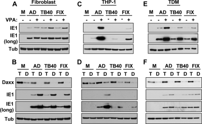
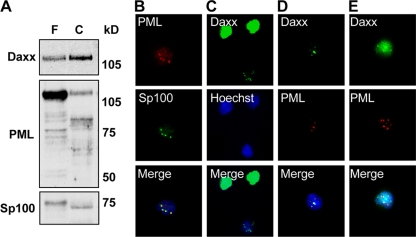
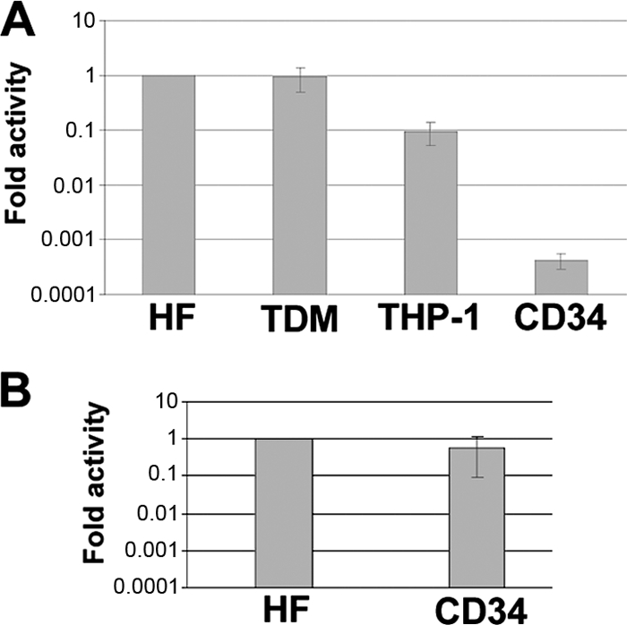
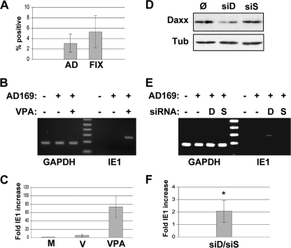
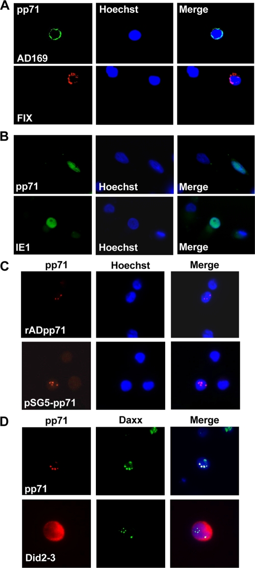
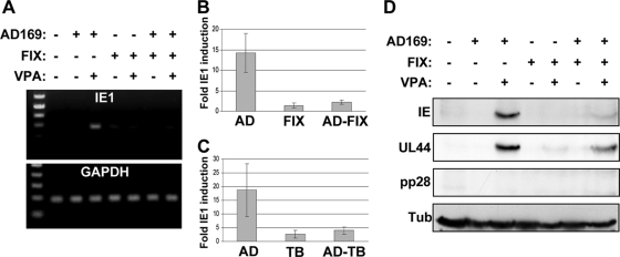
Human cytomegalovirus (HCMV) persists for the life of its host by establishing a latent infection. The identification of viral and cellular determinants of latency is the first step toward developing antiviral treatments that target and might clear or control the reservoir of latent virus. HCMV latency is established in CD34(+) cells when expression of viral immediate early (IE) proteins that initiate lytic infection is silenced. Viral IE gene expression during lytic infection is controlled by a cellular intrinsic immune defense mediated by promyelocytic leukemia nuclear body (PML-NB) proteins such as Daxx and histone deacetylases (HDACs). This defense is inactivated at the start of lytic infection by the HCMV virion tegument protein pp71, which upon viral entry traffics to the nucleus and induces Daxx degradation. Here we show that a similar defense is present, active, and not neutralized during experimental latency in CD34(+) cells infected in vitro because tegument-delivered pp71 remains in the cytoplasm. Artificial inactivation of this defense by HDAC inhibition or Daxx knockdown rescues viral IE gene expression upon infection of CD34(+) cells with a laboratory-adapted viral strain but not with clinical strains. Interestingly, coinfection of CD34(+) cells with clinical viral strains blocked the ability of an HDAC inhibitor to activate IE1 and early protein expression during infection with a laboratory-adapted strain. This suggests that in addition to the intrinsic defense, HCMV clinical strains contribute an HDAC-independent, trans-acting dominant means of control over viral gene expression during the early stages of experimental HCMV latency modeled in vitro in CD34(+) cells.


Figures

FIG. 1.
FIG. 1.
HCMV clinical isolates are susceptible to Daxx-mediated MIEP silencing. (Panel A) HFFs were mock infected (M) or treated without (−) or with (+) VPA ∼18 h prior to infection with AD169 (AD), TB40/E (TB40), or FIX at a multiplicity of infection (MOI) of 0.05. Lysates were harvested 6 h postinfection (hpi) and analyzed by Western blotting. (Panel B) TERT-HFs (T) or those constitutively expressing an shRNA to Daxx (D) were infected with AD169, TB40/E, or FIX at an MOI of 0.5. Lysates were harvested at 6 hpi and analyzed by Western blotting. (Panel C) THP-1 monocytes were mock infected or infected with AD169, TB40/E, or FIX in the absence (−) or presence (+) of VPA at an MOI of 1 for 12 h. Lysates were analyzed by Western blotting. (Panel D) THP-1 cells (T) or those expressing shRNA to Daxx (D) were infected with AD169, TB40/E, or FIX at an MOI of 1 for 24 h and analyzed by Western blotting. (Panel E) THP-1-derived macrophage (TDM) were mock infected or treated without (−) or with (+) VPA for 3 h prior to infection with AD169, TB40/E, or FIX at an MOI of 0.5. Lysates were harvested 12 hpi and analyzed by Western blotting. (Panel F) THP-1-derived macrophage (T) or those expressing shRNA to Daxx (D) were infected with AD169, TB40/E, or FIX at an MOI of 0.5 for 12 h and analyzed by Western blotting. For all experiments, tubulin (Tub) was used as a loading control. Short and long exposures are shown for IE1.
FIG. 2.
FIG. 2.
CD34+ cells contain PML-NBs. (Panel A) Fifty micrograms of protein lysate from NHDFs (F) or CD34+ cells (C) were analyzed by Western blotting. Approximate protein sizes in kDa are shown to the right. (Panel B) CD34+ cells were immobilized on coverslips and stained for PML and Sp100 (red and green, respectively). Nuclei were counterstained with Hoechst stain (blue). (Panel C) CD34+ cells stained for Daxx (green). (Panel D) CD34+ cells stained for Daxx (green) and PML (red) for which Daxx is punctate. (Panel E) CD34+ cells stained for Daxx (green) and PML (red) for which Daxx is diffuse. The magnification for all images in this figure is ×60.
FIG. 3.
FIG. 3.
The HCMV MIEP is less active in CD34+ cells than in other cell types. (Panel A) NHDF (HF), THP-1-derived macrophage (TDM), THP-1, and CD34+ cells were transduced with a recombinant adenovirus (rAD) expression system driven by the HCMV MIEP and harvested 24 h posttransduction. The relative quantity of expressed mRNA compared to input rAD genomic DNA was determined by Q-PCR (see Materials and Methods). Fold activity of the MIEP was compared to levels found in NHDFs, which were empirically set at 1. Averages and standard errors of the means for five independent experiments are shown. (Panel B) NHDF and CD34+ cells were transduced with an rAD expression system driven by the EF1α promoter and analyzed as described for panel A. Results from three independent experiments are shown, with the averages and standard errors of the means indicated.
FIG. 4.
FIG. 4.
Daxx and histone deacetylase activity control HCMV AD169 IE gene expression during infection of CD34+ cells. (Panel A) CD34+ cells were infected with AD169 (AD) or FIX at an MOI of 5 or 3, respectively. After 24 h, cells were harvested, treated with trypsin, immobilized on coverslips, and stained for pp71. The percentages of cells staining positive for pp71 are shown. Standard deviations are indicated with bars. (Panel B) CD34+ cells were mock treated (−) or treated with VPA (+) and either mock infected (−) or infected with AD169 (+) at an MOI of 1 as indicated. Cells were harvested 24 hpi, and total RNA was extracted and analyzed by RT-PCR for expression of IE1. Cellular GAPDH served as an internal control. (Panel C) Fold IE1 activation (normalized to GAPDH levels) for samples shown in panel B is shown. M, mock; V, AD169; VPA, AD169 + VPA. Bars indicate standard deviations. (Panel D) CD34+ cells were transfected without siRNA (Ø) or transfected with siRNA directed at Daxx (siD) or Skp-1 (siS). Forty-eight hours later, cells were infected with AD169 at an MOI of 1 for 24 h. Cells were harvested, and a fraction was used for Western blot analysis. Tubulin (Tub) was used as a normalization control. For the experiment shown, the Daxx level in the Daxx-knockdown cells was 42% of the untreated cells. In the Skp-1-knockdown cells, the Daxx level was 96% of the untreated cells. (Panel E) RNA was isolated from the remaining cells (treated as described for panel D above) and used in an RT-PCR assay for IE1 and GAPDH. Mock-infected (−) or AD169-infected (+) cells and cells transfected without siRNA (−), with siRNA directed at Daxx (D), or with siRNA directed at Skp-1 (S) were analyzed. (Panel F) Results from five independent experiments as outlined (panels D and E) were analyzed, and fold activation of IE1 in cells transfected with siRNA to Daxx over those transfected with siRNA to Skp-1 (siD/siS) is shown with the standard deviation (*, P value < 0.05).
FIG. 5.
FIG. 5.
Differentiation-dependent localization of tegument-delivered pp71 and IE1 expression during HCMV infection of CD34+ cells. (Panel A) CD34+ cells were infected with AD169 or FIX at an MOI of 5 or 3, respectively. After 24 h, cells were harvested, treated with trypsin, immobilized on coverslips, and stained with an antibody to pp71 (AD169, green; FIX, red). (Panel B) CD34+ cells were differentiated into dendritic cells and infected with AD169 at an MOI of 5 for 24 h. Adherent cells on coverslips were analyzed for the presence of pp71 or IE1 (both green). (Panel C) CD34+ cells were transduced with a recombinant adenovirus that expresses pp71 (rADpp71) at 1,000 particles per cell for 24 h or transfected with a pp71 expression plasmid (pSG5-pp71) for 36 h and analyzed for pp71 expression (red). (Panel D) CD34+ cells were transfected with expression plasmids for wild-type pp71 (pp71) or a pp71 mutant unable to bind Daxx (Did2-3) for 24 h and stained with antibodies to pp71 (red) and Daxx (green). In all panels, nuclei were counterstained with Hoechst stain (blue). The image magnification was ×60.
FIG. 6.
FIG. 6.
HCMV clinical isolates institute a dominant block to IE gene expression. (Panel A) CD34+ cells were mock infected (−), infected (+) with AD169 (MOI of 1) or FIX (MOI of 1), or coinfected with both (each at an MOI of 1 added to the cells simultaneously [total MOI of 2]) in the absence (−) or presence (+) of VPA. Cells were harvested 24 hpi, and isolated RNA was used in an RT-PCR for IE1 and GAPDH expression. (Panel B) Fold activation of IE1 during infection in the presence of VPA compared to virus alone was determined for experiments described for panel A. (Panel C) Experiments were conducted as outlined above by using AD169 (AD; MOI of 1), clinical isolate TB40/E (TB; MOI of 1), or both (AD-TB; each at an MOI of 1, added to the cells simultaneously [total MOI of 2]). Fold IE1 activation with virus plus VPA over virus alone is shown. For panels B and C, results from four (B) or three (C) independent experiments with standard errors of the means shown. (Panel D) CD34+ cells left untreated (−) or treated at the time of infection with 1 mM VPA (+) as indicated were infected with AD169 (MOI of 1) or FIX (MOI of 1) or coinfected with a mixture of AD169 and FIX (each at an MOI of 1 for a total MOI of 2) for 24 h, after which the cells were washed free of virus and VPA and cultured for an additional 6 days. Equal cell numbers were lysed in RIPA buffer, and their protein contents were analyzed by Western blotting with the indicated antibodies. Tubulin was used as a loading control. Note that titers are based on fibroblast infection and do not represent the efficiency with which HCMV enters CD34+ cells (Fig. 4A).

Similar articles

Cited by

References

    1. Bego, M., J. Maciejewski, S. Khaiboullina, G. Pari, and S. St. Jeor. 2005. Characterization of an antisense transcript spanning the UL81-82 locus of human cytomegalovirus. J. Virol. 79:11022-11034. - PMC - PubMed
    1. Cantrell, S. R., and W. A. Bresnahan. 2006. Human cytomegalovirus (HCMV) UL82 gene product (pp71) relieves hDaxx-mediated repression of HCMV replication. J. Virol. 80:6188-6191. - PMC - PubMed
    1. Cha, T. A., E. Tom, G. W. Kemble, G. M. Duke, E. S. Mocarski, and R. R. Spaete. 1996. Human cytomegalovirus clinical isolates carry at least 19 genes not found in laboratory strains. J. Virol. 70:78-83. - PMC - PubMed
    1. Cheung, A. K., A. Abendroth, A. L. Cunningham, and B. Slobedman. 2006. Viral gene expression during the establishment of human cytomegalovirus latent infection in myeloid progenitor cells. Blood 108:3691-3699. - PubMed
    1. Cheung, A. K., D. J. Gottlieb, B. Plachter, S. Pepperl-Klindworth, S. Avdic, A. L. Cunningham, A. Abendroth, and B. Slobedman. 2009. The role of the human cytomegalovirus UL111A gene in down-regulating CD4+ T-cell recognition of latently infected cells: implications for virus elimination during latency. Blood 114:4128-4137. - PubMed

Publication types

MeSH terms