. 2010 Jun;84(11):5775-89.
doi: 10.1128/JVI.02343-09. Epub 2010 Mar 24.

Role of annexin A2 in the production of infectious hepatitis C virus particles

Affiliations

Role of annexin A2 in the production of infectious hepatitis C virus particles

Perdita Backes et al. J Virol. 2010 Jun.

Abstract

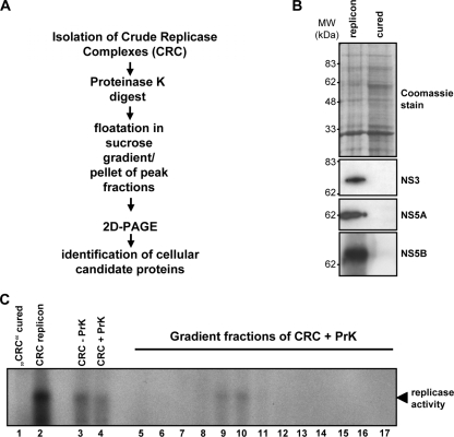
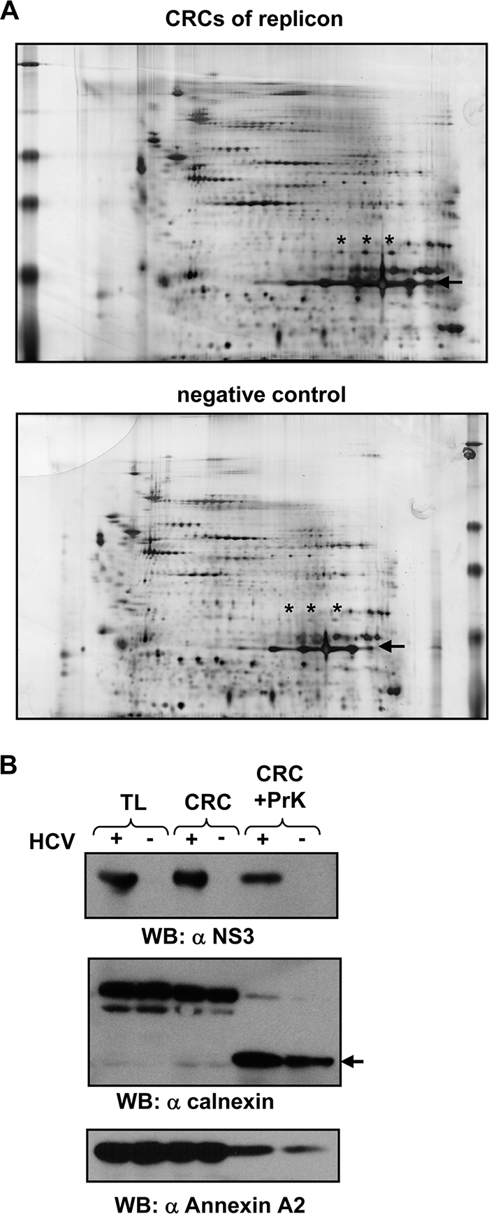
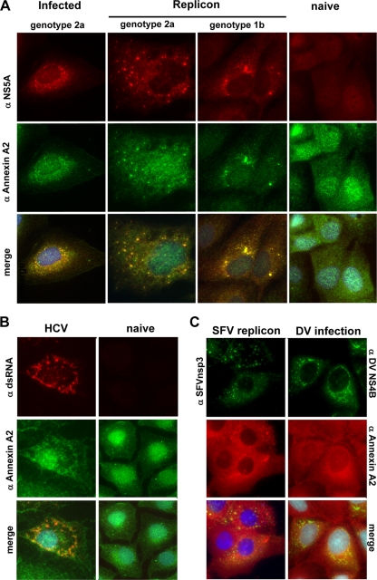
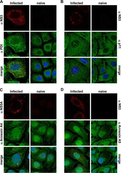
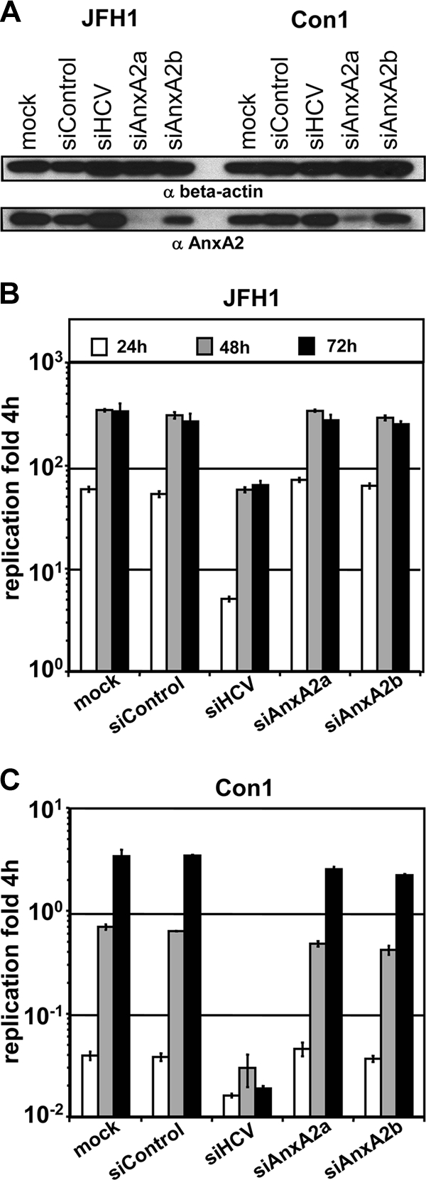
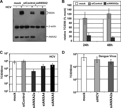
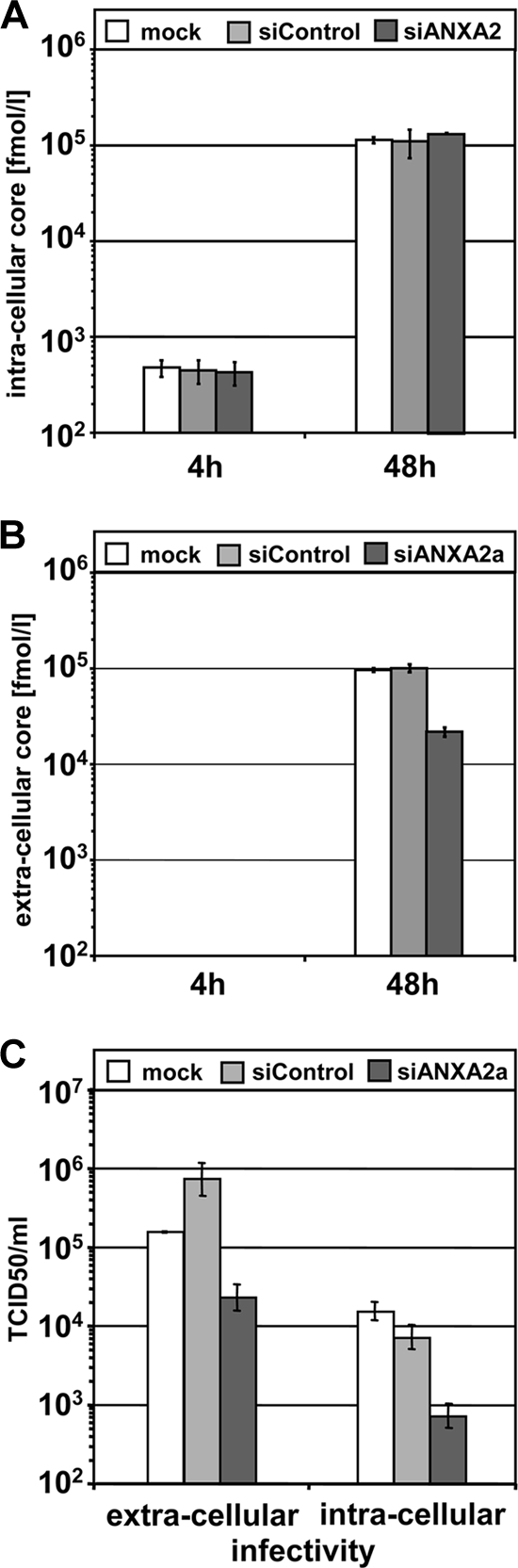
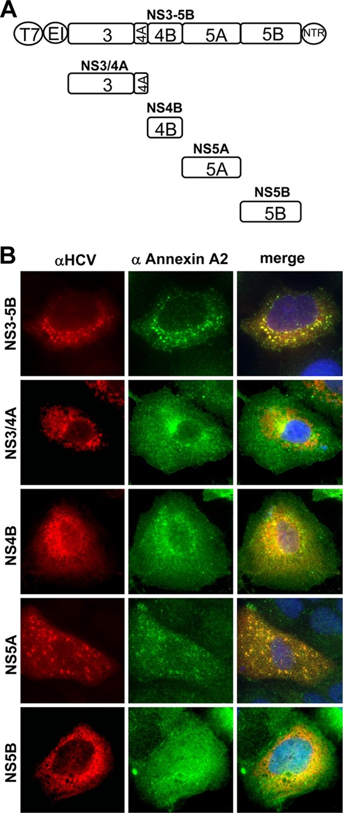
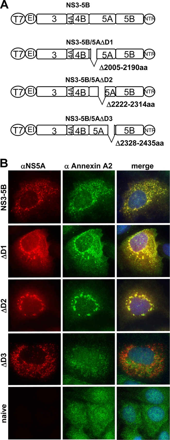
Hepatitis C virus (HCV) is an important human pathogen affecting 170 million chronically infected individuals. In search for cellular proteins involved in HCV replication, we have developed a purification strategy for viral replication complexes and identified annexin A2 (ANXA2) as an associated host factor. ANXA2 colocalized with viral nonstructural proteins in cells harboring genotype 1 or 2 replicons as well as in infected cells. In contrast, we found no obvious colocalization of ANXA2 with replication sites of other positive-strand RNA viruses. The silencing of ANXA2 expression showed no effect on viral RNA replication but resulted in a significant reduction of extra- and intracellular virus titers. Therefore, it seems likely that ANXA2 plays a role in HCV assembly rather than in genome replication or virion release. Colocalization studies with individually expressed HCV nonstructural proteins indicated that NS5A specifically recruits ANXA2, probably by an indirect mechanism. By the deletion of individual NS5A subdomains, we identified domain III (DIII) as being responsible for ANXA2 recruitment. These data identify ANXA2 as a novel host factor contributing, with NS5A, to the formation of infectious HCV particles.


Figures

FIG. 1.
FIG. 1.
Establishment of a purification scheme for the proteomic analysis of HCV replication complexes. (A) Schematic representation of a sample preparation for the proteomic analysis of cell extracts from HCV replicon and cured replicon cells. (B) Analysis of cell extracts from replicon and cured replicon cells. Identical portions of the pellet collected from the peak gradient fractions were subjected to SDS-PAGE. Proteins included in this fraction were either stained with Coomassie brilliant blue (top) or analyzed by immunoblotting using a polyclonal antiserum specific for HCV proteins (given in the right of each panel). (C) Replicase activity of sucrose gradient fractions of CRCs treated with proteinase K (PrK). Gradient fractions were collected from the top, and 4 μl of each gradient fraction as well as nontreated (−PrK) and PrK-treated (+PrK) CRCs were analyzed for in vitro replicase activity. A concentrated CRC fraction from replicon cells served as the positive control, and a corresponding fraction from cured cells served as the negative control.
FIG. 2.
FIG. 2.
Identification of annexin A2 as a potential component of HCV genotype 1b replication complexes and evaluation by the Western blot analysis (WB) of cell extracts from genotype 2a replicon cells. (A) 2D-PAGE of membrane fractions from cells harboring subgenomic replicons or their cured counterparts treated according to the scheme in Fig. 1A. Proteinase K is marked by the arrow, and ANXA2 spots are marked with asterisks. (B) Detection of viral and cellular proteins in CRCs before and after proteinase K digestion. Viral and cellular antigens were detected in total-lysate (TL), CRC, and digested CRC fractions (CRC+PrK) of cells harboring a subgenomic genotype 2a replicon (isolate JFH1) and their cured counterparts. The viral protein was detected with a polyclonal antiserum directed against the helicase domain of NS3 (α-NS3). The band corresponding to C-terminally truncated calnexin is marked by an arrow. ANXA2 was stained with a monoclonal mouse antibody. Note that 5-fold-larger amounts of the proteinase K-treated CRC fractions were loaded compared to those of untreated samples.
FIG. 3.
FIG. 3.
Subcellular localization of ANXA2. (A) Colocalization of NS5A with annexin A2 in infected Huh7.5 or cell lines harboring persistent genotype 1b (Con1, clone 9-13) or genotype 2a (JFH1) replicons. Cells containing subgenomic replicons and naïve Huh-7 cells were subjected to immunofluorescence analysis 48 h after seeding. Huh7.5 cells were analyzed 48 h after infection. ANXA2 was detected by a polyclonal rabbit antiserum and a secondary goat anti-rabbit antibody labeled with Alexa 488 (middle row). NS5A was detected with monoclonal antibody 9E10, recognizing genotype 1b and 2a subtypes, and a secondary goat anti-mouse antibody labeled with Alexa 546 (upper row). Naïve Huh-7 cells served as the negative control (Huh-7) and were processed in parallel. (B) Colocalization of ANXA2 and double-stranded RNA in HCV JFH1 replicon cells. Cells containing subgenomic replicons and naïve Huh-7 cells were subjected to immunofluorescence analysis 48 h after seeding. ANXA2 was detected by a polyclonal rabbit antiserum and a secondary goat anti-rabbit antibody labeled with Alexa 488. dsRNA was detected with monoclonal mouse antibody J2 and a secondary goat anti-mouse antibody labeled with Alexa 546. (C) Localization of annexin A2 and the replication complexes of Semliki Forest virus (SFV) in BHK-21 cells or dengue virus (DV) in Huh-7 cells. Cells were fixed 24 h posttransfection or infection and stained with monoclonal mouse ANXA2 antibody HH7 and a secondary goat anti-mouse antibody labeled with Alexa 546. Viral antigens were detected with polyclonal antisera directed against SFV nsp3 or DV NS4B, respectively, and a goat anti-rabbit antibody labeled with Alexa 488.
FIG. 4.
FIG. 4.
Localization of HCV nonstructural proteins and different cellular proteins in Huh7-Lunet/CD81 cells infected with HCV JC1. Infected cells (left rows) or naive Huh7-Lunet/CD81 cells (right rows) were subjected to immunofluorescence analysis 48 h after seeding. Subcellular localization of PDI (middle row) and NS3 (top row) (A), p11 (S100/A10, middle row) and NS3 (top row) (B), annexin A4 (middle row) and NS5A (top row) (C), and annexin A5 (middle row) and NS3 (top row) (D). Note that NS3 and NS5A almost perfectly colocalize in HCV infected cells.
FIG. 5.
FIG. 5.
Effect of annexin A2 knockdown on replication of subgenomic reporter replicons. Huh7-Lunet cells were transfected with the indicated siRNAs, and 3 days later they were electroporated with a second dose of the same siRNA together with JFH1 or Con1 subgenomic replicons encoding firefly luciferase and analyzed for ANXA2 knockdown efficiency (A) and HCV replication (B and C). (A) Knockdown efficiency of ANXA2 siRNAs analyzed by Western blotting. Total cell lysates were harvested 48 h after electroporation with the indicated siRNAs and HCV replicon RNAs and subjected to Western blot analysis. β-Actin (upper) as well as ANXA2 (lower) were stained with monoclonal mouse antibodies. (B and C) The replication efficiency of HCV genotype 1b (B) or 2a (C) replicons was determined by luciferase reporter assay. Luciferase activity is expressed as the ratio of relative light units (RLU) obtained at 24 (white bars), 48 (gray bars), and 72 h (black bars) relative to the luciferase activity 4 h after electroporation to normalize for transfection efficiency. Mean values and standard deviations from a representative experiment out of five independent experiments are shown.
FIG. 6.
FIG. 6.
Impact of the silencing of ANXA2 and other annexins on HCV or dengue virus titers. Huh7-Lunet cells were transfected with the indicated siRNAs, electroporated with a second dose of the same siRNA together with viral genomes, and analyzed for ANXA2 knockdown efficiency (A) and titers of HCV (B and C) or dengue virus (D). (A) Reduction of ANXA2 levels after knockdown. Shown is a Western blot of cell lysates harvested 48 h postelectroporation of Huh7-Lunet cells with HCV JC1 in vitro transcripts and the indicated siRNAs. Antigens were detected by monoclonal mouse antibodies directed against NS5A, β-actin, and ANXA2 as indicated on the right. (B) ANXA2 knockdown impairs the production of infectious HCV. The relative infectivity in supernatants at 24 and 48 h after electroporation with JC1 and the indicated siRNA is shown. Mean values and standard deviations of viral titers from seven independent experiments are given in percentages normalized to TCID50 titers in mock-silenced cells. (C) No impact of ANXA4 or ANXA5 knockdown on viral titers. HCV infectivity titers in supernatants harvested 48 h after the electroporation of cells with HCV JC1 full-length RNA and the indicated siRNAs are shown. The TCID50/ml of a representative experiment are shown. Knockdown efficiency was checked by Western blotting (not shown). (D) No reduction of dengue virus titers upon ANXA2 silencing. Titers of dengue virus genotype 2 in supernatants harvested 48 h after the electroporation of presilenced cells with DV full-length RNA and siRNAs shown as the TCID50/ml of a representative experiment. An siRNA directed against HCV was used as a negative control in this experiment.
FIG. 7.
FIG. 7.
Extra- and intracellular amounts of viral Core protein and infectivity titers after ANXA2 knockdown. Huh7-Lunet cells were transfected with the indicated siRNAs, electroporated with a second dose of the same siRNA together with HCV JC1 full-length viral genome, and analyzed for intra- and extracellular Core protein and TCID50. (A) Intracellular Core levels (fmol/liter) in freeze-thaw lysates of silenced cells harvested 4 and 48 h after electroporation with the indicated siRNAs and HCV full-length RNA. (B) Extracellular Core levels (fmol/liter) in supernatants of cells 4 and 48 h after electroporation. (C) Extra- and intracellular infectivity titers in freeze-thaw lysates and supernatants of cells harvested 48 h after electroporation. Mean values and standard deviations from one out of three (A and B) or five (C) representative experiments are shown.
FIG. 8.
FIG. 8.
Localization of annexin A2 and HCV antigens in cells expressing individual nonstructural proteins (genotype 2a). (A) Schematic representation of expression constructs used in this experiment. Viral nonstructural proteins are represented as squares; the T7 promoter (T7), EMCV IRES (EI), and 3′ nontranslated regions (NTR) are depicted as circles. The individual NS proteins are shown in relation to the NS3-5B polyprotein. (B) Lunet T7 cells were transfected 24 h after seeding with pTM constructs encoding viral proteins as indicated on the left and subjected to immunofluorescence analysis 24 h after transfection either using primary antibodies detecting HCV antigens and secondary antibodies labeled with Alexa 546 (left) or primary antibodies detecting ANXA2 and secondary antibodies labeled with Alexa 488 (middle). Cells expressing NS3/4A, 4B, and 5B were stained with polyclonal antisera against the individual viral proteins and monoclonal anti-ANXA2 antibody HH7. In cells expressing NS3-5B or NS5A, the viral antigen was detected by mouse monoclonal anti-NS5A antibody 9E10 and ANXA2 with a polyclonal antiserum.
FIG. 9.
FIG. 9.
Subcellular localization of annexin A2 and NS5A after expression of NS3 to NS5B coding sequences (genotype 2a) harboring deletions of individual NS5A subdomains. (A) Schematic representation of expression constructs used in this experiment. Internal deletions of NS5A are marked by gaps at the relevant positions; numbers next to the gap refer to the amino acid position of the deletion. Note that the size of the deletions is not drawn to scale. For further explanation, see the legend to Fig. 8A. (B) Lunet T7 cells were transfected 24 h after seeding with pTM constructs encoding viral proteins as indicated on the left and subjected to immunofluorescence analysis 24 h after transfection either using primary antibodies detecting HCV NS5A and secondary antibodies labeled with Alexa 546 (left) or a polyclonal antiserum detecting ANXA2 and secondary antibodies labeled with Alexa 488 (middle). NS5A was stained by monoclonal mouse antibody 9E10 or 2F6 in the case of ΔDIII, since DIII contains the epitope detected by 9E10.

References

    1. Ali, N., K. D. Tardif, and A. Siddiqui. 2002. Cell-free replication of the hepatitis C virus subgenomic replicon. J. Virol. 76:12001-12007. - PMC - PubMed
    1. Alter, M. J. 2007. Epidemiology of hepatitis C virus infection. World J. Gastroenterol. 13:2436-2441. - PMC - PubMed
    1. Appel, N., M. Zayas, S. Miller, J. Krijnse-Locker, T. Schaller, P. Friebe, S. Kallis, U. Engel, and R. Bartenschlager. 2008. Essential role of domain III of nonstructural protein 5A for hepatitis C virus infectious particle assembly. PLoS Pathog. 4:e1000035. - PMC - PubMed
    1. Bartenschlager, R., M. Frese, and T. Pietschmann. 2004. Novel insights into hepatitis C virus replication and persistence. Adv. Virus Res. 63:71-180. - PubMed
    1. Beaton, A. R., J. Rodriguez, Y. K. Reddy, and P. Roy. 2002. The membrane trafficking protein calpactin forms a complex with bluetongue virus protein NS3 and mediates virus release. Proc. Natl. Acad. Sci. USA 99:13154-13159. - PMC - PubMed

Publication types

LinkOut - more resources