. 2010 May;137(9):1421-30.
doi: 10.1242/dev.048678. Epub 2010 Mar 24.

Recombineering Hunchback identifies two conserved domains required to maintain neuroblast competence and specify early-born neuronal identity

Affiliations

Recombineering Hunchback identifies two conserved domains required to maintain neuroblast competence and specify early-born neuronal identity

Khoa D Tran et al. Development. 2010 May.

Abstract

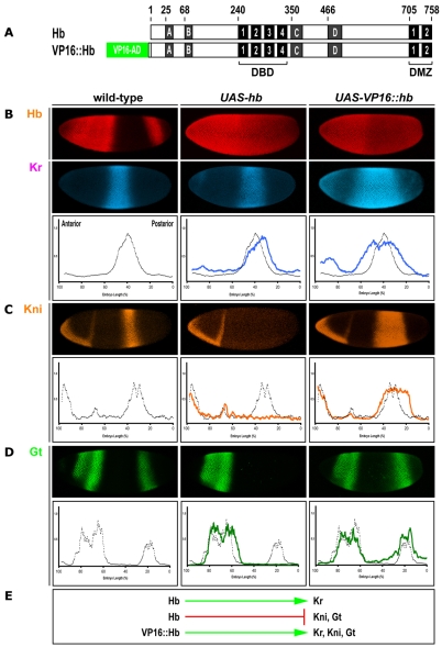
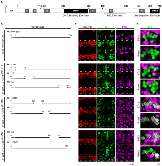
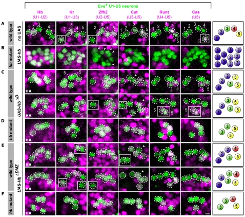
The Hunchback/Ikaros family of zinc-finger transcription factors is essential for specifying the anterior/posterior body axis in insects, the fate of early-born pioneer neurons in Drosophila, and for retinal and immune development in mammals. Hunchback/Ikaros proteins can directly activate or repress target gene transcription during early insect development, but their mode of action during neural development is unknown. Here, we use recombineering to generate a series of Hunchback domain deletion variants and assay their function during neurogenesis in the absence of endogenous Hunchback. Previous studies have shown that Hunchback can specify early-born neuronal identity and maintain 'young' neural progenitor (neuroblast) competence. We identify two conserved domains required for Hunchback-mediated transcriptional repression, and show that transcriptional repression is necessary and sufficient to induce early-born neuronal identity and maintain neuroblast competence. We identify pdm2 as a direct target gene that must be repressed to maintain competence, but show that additional genes must also be repressed. We propose that Hunchback maintains early neuroblast competence by silencing a suite of late-expressed genes.


Figures

Fig. 1.
Fig. 1.
VP16::Hb acts as a constitutive transcriptional activator. (A) Schematic of wild-type Hb protein and the VP16::Hb protein chimera showing the previously characterized conserved domains. DBD, DNA-binding domain; DMZ, dimerization domain. (B-D) Expression of gap genes in the Drosophila embryonic blastoderm in wild-type, v32a-gal4 UAS-hb and v32a-gal4 UAS-VP16::hb embryos. (E) Summary of gene interactions from B-D.
Fig. 2.
Fig. 2.
VP16::Hb activates Hb direct and indirect targets in the CNS. (A-C) VP16::Hb activates endogenous hb. In situ hybridization against endogenous hb mRNA. Each panel shows the neuroblast layer of a stage 12 Drosophila embryo with the anterior to the left. Lines in B and C indicate the engrailed-gal4 expression domain. (D-F) VP16::Hb activates the direct target pdm. Histochemical detection of Pdm (brown) and the segment boundary marker Engrailed (purple). Each panel shows the neuroblast layer of a stage 15 embryo with anterior to the left. (G-I) Expression of Kr, zfh2, cut, runt and cas in wild-type, engrailed-gal4 UAS-hb and engrailed-gal4 UAS-VP16::hb, hb mutant embryos. Each panel shows a two-dimensional projection of approximately two segments of the ventral nerve cord of a stage 16 embryo. Lines as in B,C. Anterior is up. (J) Summary of gene interactions in the CNS from A-I.
Fig. 3.
Fig. 3.
Hb maintains early neuroblast competence through the repression of multiple target genes. (A-A‴) Quantification of U neurons (within dashed box) specified in various genetic backgrounds. Wild type, average of 5; engrailed-gal4 UAS-hb, hb mutant, average of 12±2 (s.d.), range 6-18; engrailed-gal4 UAS-VP16::hb, hb mutant, average of 6±2, range 2-12. P⪡0.001 for all experiments. (B,C) Neuroblast expression profile at early stage 12. One hemisegment is shown. Dashed circles outline neuroblasts, and solid lines indicate the engrailed-gal4 expression domain. Hb activates Kr and represses pdm and cas in neuroblasts, whereas VP16::Hb activates all three. (D,D′) Co-misexpression of Hb + Pdm and Hb + Cas results in fewer ectopic U neurons. UAS-hb UAS-HA control, average of 17±2, range 13-22; UAS-hb UAS-pdm2, average of 9±2, range 3-14; UAS-hb UAS-cas, average of 15±2, range 8-20. P⪡0.001 for all experiments. (E) Overexpression of VP16::Hb in a pdm mutant embryo does not result in the recovery of ectopic U neurons. pros-gal4 UAS-hb, average of 9±2.5, range 5-16; pros-gal4 UAS-VP16::hb, average of 5.6±1, range 4-9; pros-gal4 UAS-VP16::hb, pdm mutant, average of 4.3±1, range 2-8. P⪡0.001 for all experiments. (F) Summary and comparison of the competence windows generated by Hb and VP16::Hb.
Fig. 4.
Fig. 4.
The Hunchback D and DMZ domains are required for transcriptional repression and maintenance of neuroblast competence. (A) Schematic of Drosophila Hb protein showing conserved domains and deletion breakpoints (bold). (B) Deletions grouped by functional phenotype. (C) In each set of panels, which refer to the corresponding deletions in B, two segments of a stage 11 embryo are shown with neuroblasts stained for Kr and Pdm proteins. Dashed lines indicate engrailed-gal4 expression domains. Wild-type Hb, HbΔAB, HbΔB′ and HbΔE can activate Kr and repress pdm; HbΔDBD and HbΔC are non-functional in the CNS; HbΔD and HbΔDMZ can activate Kr, but cannot repress pdm. (D)U neurons (encircled) specified by Hb and each deletion construct.
Fig. 5.
Fig. 5.
The Hb D and DMZ domains are required for the first temporal identity. Each panel shows a two-dimensional projection of U neurons from one hemisegment of a stage 16 Drosophila embryo; medial is to the left and anterior is to the top. The U1-U5 neurons (as illustrated to the right) can be uniquely identified based on the indicated molecular markers. For quantification of U neuron identity, see Table 1. Scale bar: 3 μm. (A) Wild-type embryo. (B) engrailed-gal4 UAS-hb, hb mutant embryo. Ectopic early-born U1/U2 neurons are specified. Arrowheads indicate weak Zfh2+ cells. (C) engrailed-gal4 UAS-hbΔD embryo. An ectopic U2 or U3 neuron is found (arrowhead) in 50% of hemisegments. All hemisegments contain two Cas+ U5 neurons. (D) engrailed-gal4 UAS-hbΔD in a hb mutant embryo. Most hemisegments contain an ectopic U3 neuron but no U1 or U2 neurons are specified. (E) engrailed-gal4 UAS-hbΔDMZ embryo. An ectopic U2 or U3 neuron (arrowheads) is found in 25% of hemisegments. All other U neurons differentiate as in wild type. (F) engrailed-gal4 UAS-hbΔDMZ in a hb mutant embryo. Most hemisegments contain an ectopic U3 neuron and no U1 or U2 neurons are specified.
Fig. 6.
Fig. 6.
Models for Hb-mediated transcriptional regulation of neuroblast competence in Drosophila. Proposed molecular interactions underlying Hb function in the CNS. (A) Hb binds to its consensus sequence and recruits co-activators (and/or outcompetes repressors) to promote gene expression. (B) Hb monomers bind to genomic DNA and recruit repressor complexes. (C) Hb monomer binds to the regulatory region of a target gene, which is then repressed by dimerization with a second Hb monomer bound within a heterochromatin domain. (D) Hb dimerization is required for the recruitment of repressor complexes, which might include Mi2. Magenta ovals, Hb; smaller ovals, Hb dimerization domain; magenta squares, Hb binding sites; dark-green line, genomic DNA; green arrow, transcription start site; dark-green boxes, gene; A, activator; green square, activator binding sites; R, repressor; red squares, repressor binding sites.

Similar articles

Cited by

References

    1. Albertson R., Chabu C., Sheehan A., Doe C. Q. (2004). Scribble protein domain mapping reveals a multistep localization mechanism and domains necessary for establishing cortical polarity. J. Cell Sci. 117, 6061-6070 - PubMed
    1. Baumgardt M., Karlsson D., Terriente J., Diaz-Benjumea F. J., Thor S. (2009). Neuronal subtype specification within a lineage by opposing temporal feed-forward loops. Cell 139, 969-982 - PubMed
    1. Berman B. P., Nibu Y., Pfeiffer B. D., Tomancak P., Celniker S. E., Levine M., Rubin G. M., Eisen M. B. (2002). Exploiting transcription factor binding site clustering to identify cis-regulatory modules involved in pattern formation in the Drosophila genome. Proc. Natl. Acad. Sci. USA 99, 757-762 - PMC - PubMed
    1. Berman B. P., Pfeiffer B. D., Laverty T. R., Salzberg S. L., Rubin G. M., Eisen M. B., Celniker S. E. (2004). Computational identification of developmental enhancers: conservation and function of transcription factor binding-site clusters in Drosophila melanogaster and Drosophila pseudoobscura. Genome Biol. 5, R61 - PMC - PubMed
    1. Bischof J., Maeda R. K., Hediger M., Karch F., Basler K. (2007). An optimized transgenesis system for Drosophila using germ-line-specific phiC31 integrases. Proc. Natl. Acad. Sci. USA 104, 3312-3317 - PMC - PubMed

Publication types

MeSH terms