. 2010 May;137(9):1431-9.
doi: 10.1242/dev.045427. Epub 2010 Mar 24.

Abnormal hair follicle development and altered cell fate of follicular keratinocytes in transgenic mice expressing DeltaNp63alpha

Affiliations

Abnormal hair follicle development and altered cell fate of follicular keratinocytes in transgenic mice expressing DeltaNp63alpha

Rose-Anne Romano et al. Development. 2010 May.

Erratum in

  • Development. 2010 May;137(10):1775

Abstract

The transcription factor p63 plays an essential role in epidermal morphogenesis. Animals lacking p63 fail to form many ectodermal organs, including the skin and hair follicles. Although the indispensable role of p63 in stratified epithelial skin development is well established, relatively little is known about this transcriptional regulator in directing hair follicle morphogenesis. Here, using specific antibodies, we have established the expression pattern of DeltaNp63 in hair follicle development and cycling. DeltaNp63 is expressed in the developing hair placode, whereas in mature hair its expression is restricted to the outer root sheath (ORS), matrix cells and to the stem cells of the hair follicle bulge. To investigate the role of DeltaNp63 in hair follicle morphogenesis and cycling, we have utilized a Tet-inducible mouse model system with targeted expression of this isoform to the ORS of the hair follicle. DeltaNp63 transgenic animals display dramatic defects in hair follicle development and cycling, eventually leading to severe hair loss. Strikingly, expression of DeltaNp63 leads to a switch in cell fate of hair follicle keratinocytes, causing them to adopt an interfollicular epidermal (IFE) cell identity. Moreover, DeltaNp63 transgenic animals exhibit a depleted hair follicle stem-cell niche, which further contributes to the overall cycling defects observed in the mutant animals. Finally, global transcriptome analysis of transgenic skin identified altered expression levels of crucial mediators of hair morphogenesis, including key members of the Wnt/beta-catenin signaling pathway, which, in part, account for these effects. Our data provide evidence supporting a role for DeltaNp63alpha in actively suppressing hair follicle differentiation and directing IFE cell lineage commitment.


Figures

Fig. 1.
Fig. 1.
ΔNp63 expression analysis during hair follicle development and cycling. (A,B) At E14.5, expression of ΔNp63 protein can be detected in the developing placodes and continues to be expressed in the down-growing hair germ at E16.5 (B). Dotted lines indicate the epidermal-dermal junction. (C) By postnatal day 1 (P1), ΔNp63 is expressed along the entire length of the outer root sheath (ORS) as well as the matrix cells. (D) At P21 (telogen), ΔNp63 expression is localized to the basal layer, ORS and the bulge region (brackets, insets) of the hair follicle and secondary hair germ (Hg). (E,F) Co-staining with K15 and Sox9 reveals co-localization of ΔNp63 to hair follicle stem cells. Scale bars: 50μm.
Fig. 2.
Fig. 2.
Gross morphology and histological analysis of ΔNp63α transgenic mice. (A) Gross morphology reveals that the BG animals have thickened scaly skin and fail to grow hair as compared with control littermates at P16. (B) Western blot analysis using skin whole-cell extracts reveals that BG animals express the transgene (α-HA) as well as increased levels of ΔNp63 (α-ΔNp63α) as compared with wild-type control littermates at P16. β-tubulin serves as a loading control. (C) Dorsal skin sections from various developmental time-points stained with Hematoxylin and Eosin (H&E). At newborn (NB), BG animals are indistinguishable from their control littermates (top panel). At P16, the BG interfollicular epidermis (IFE) is hyperplastic and displays a reduced granular layer (middle panel inset). Hair follicles from the BG animals (middle panel) are enlarged (yellow arrow) and have increased amounts of keratinized tissue localized to the hair shaft region (black arrows). By P28, when wild-type hair follicles are in telogen, the BG follicles continue to grow into the dermis (arrow, lower panel), suggesting a failure of these follicles to cycle properly. Scale bars: 75μm in upper and middle panels; 100 μm in lower panels.
Fig. 3.
Fig. 3.
Analysis of the interfollicular epidermis. (A-F) Immunofluorescence staining of dorsal skin sections using antibodies against K5, K1 and K10 shows an expansion of the proliferating basal layer (K5) and spinous layer (K1/K10) in the IFE of the BG animals. (G,H) Conversely, there is a dramatic reduction in loricrin expression in the BG animals. (I-L) Evaluation of involucrin and filaggrin expression demonstrates increased expression levels in the BG IFE as compared with wild-type animals. Dotted lines indicate the epidermal-dermal junction. Scale bar: 75μm.
Fig. 4.
Fig. 4.
Alterations in the hair follicle differentiation program of ΔNp63αBG animals. (A-R) Immunofluorescence staining used to detect changes in the hair follicle differentiation program of ΔNp63αBG animals at P13. Antibodies used for each staining are shown in lower left panels. Scale bars: 100μm in C-H; 75μm in A,B,I-L,O,P; 60 μm in Q,R; 50 μm in M,N.
Fig. 5.
Fig. 5.
Reduced matrix cell proliferation in the hair follicles of ΔNp63αBG animals. Dorsal skin sections from P13 were stained with Ki67 (upper panel) and PCNA (lower panel). Matrix cells of BG hair follicles show reduced numbers of proliferating cells as compared with control littermates (arrows). Scale bar: 75 μm.
Fig. 6.
Fig. 6.
Follicular keratinocyte transformation in BG animals. Overexpression of ΔNp63α results in the transition of hair follicle keratinocytes to adopt an IFE cell fate. Dorsal skin sections from P28 were stained with K1 and filaggrin, markers of the IFE. Compared with wild-type hair follicles, BG hair follicles express K1 and filaggrin. A higher magnification is shown in the insets. Arrows indicate cells expressing both K14/K1 or K14/Fil respectively. All sections are stained with K14 (red), K1 or Fil (green), and DAPI (blue). Scale bar: 75μm.
Fig. 7.
Fig. 7.
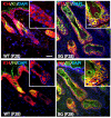
Forced expression of ΔNp63α results in hair follicle stem cell depletion. Dorsal skin sections from P21 were stained with various stem cell markers of the hair follicle bulge. As compared with wild-type control hair follicles, follicles of the BG animal show a complete loss of K15 (green), Sox9 (red) and S100A6 (red) expression, suggesting a depletion of hair follicle stem cells of the bulge. A higher magnification is shown in the insets. Scale bar: 100 μm.
Fig. 8.
Fig. 8.
Altered hair-follicle-specific genes and signaling pathways in ΔNp63α mutant animals. Heat map representation of microarray data demonstrating a downregulation of genes involved hair shaft and inner root sheath (IRS) development, genes involved in hair follicle stem cell maintenance and various signaling pathways in ΔNp63α animals. The colour scale represents the expression level of a gene above (red), below (green), or at the mean expression level (black) across all samples.
Fig. 9.
Fig. 9.
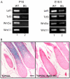
Loss of Wnt/β-catenin signaling in ΔNp63α animals. (A)RT-PCR analysis of mRNA transcripts from wild-type and BG animals. Semi-quantitative RT-PCR reveals a dramatic downregulation in the levels of several genes belonging to the Wnt/β-catenin signaling pathway at P16 and E18.5. (B) TOPGAL and TOPGALΔNp63αBG dorsal skin sections at P13 were stained for lacZ expression and counter-stained with Eosin. Staining reveals Wnt/β-catenin activity in the lower portion of the hair shaft and matrix region of the TOPGAL animals (left panel). lacZ expression in TOPGALΔNp63αBG hair follicles is dramatically reduced (right panel). Scale bar: 50μm.

Similar articles

Cited by

References

    1. Andl T., Reddy S. T., Gaddapara T., Millar S. E. (2002). WNT signals are required for the initiation of hair follicle development. Dev. Cell 2, 643-653 - PubMed
    1. Barbieri C. E., Pietenpol J. A. (2006). p63 and epithelial biology. Exp. Cell Res. 312, 695-706 - PubMed
    1. Blanpain C., Fuchs E. (2007). p63: revving up epithelial stem-cell potential. Nat. Cell Biol. 9, 731-733 - PubMed
    1. Callahan C. A., Oro A. E. (2001). Monstrous attempts at adnexogenesis: regulating hair follicle progenitors through Sonic hedgehog signaling. Curr. Opin. Genet. Dev. 11, 541-546 - PubMed
    1. Candi E., Rufini A., Terrinoni A., Dinsdale D., Ranalli M., Paradisi A., De Laurenzi V., Spagnoli L. G., Catani M. V., Ramadan S., et al. (2006). Differential roles of p63 isoforms in epidermal development: selective genetic complementation in p63 null mice. Cell Death Differ. 13, 1037-1047 - PubMed

Publication types