Intratendinous ganglion cyst of the semimembranosus tendon
- PMID: 20335437
- PMCID: PMC3473447
- DOI: 10.1259/bjr/23178227
Intratendinous ganglion cyst of the semimembranosus tendon
Abstract
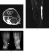
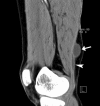
Intratendinous ganglion cyst is a very rare lesion with an unknown aetiology that originates within the tendon. We encountered a case of 43-year-old woman who complained of a palpable, non-tender mass in the thigh with increasing swelling. An intratendinous ganglion cyst in the semimembranosus tendon of the lower extremity was diagnosed and located by ultrasound and MRI. Nine months after a surgical excision, there were recurrent ganglion cysts along the semimembranosus tendon. We describe this case with a review of the relevant literature.
Figures


References
-
- McCarthy CL, McNally EG. The MRI appearance of cystic lesions around the knee. Skeletal Radiol 2004;33:187–209 - PubMed
-
- Costa CR, Morrison WB, Carrino JA, Raiken SM. MRI of an intratendinous ganglion cyst of the peroneus brevis tendon. AJR Am J Roentgenol 2003;181:890–1 - PubMed
-
- Rayan GM. Intratendinous ganglion. A case report. Orthop Rev 1989;18:449–51 - PubMed
-
- Ikeda K, Tomita K, Matsumoto H. Intratendinous ganglion in the extensor tendon of a finger: A case report. J Orthop Surg (Hong Kong) 2001;9:63–5 - PubMed
-
- Steiner E, Steinbach LS, Schnarkowski P, Tirman PF, Genant HK. Ganglia and cysts around joints. Radiol Clin North Am 1996;34:395–425, xi–xii. - PubMed

