. 2010 Mar 24;30(12):4246-55.
doi: 10.1523/JNEUROSCI.3351-09.2010.

Astrocyte-produced ephrins inhibit schwann cell migration via VAV2 signaling

Affiliations

Astrocyte-produced ephrins inhibit schwann cell migration via VAV2 signaling

Fardad T Afshari et al. J Neurosci. .

Abstract

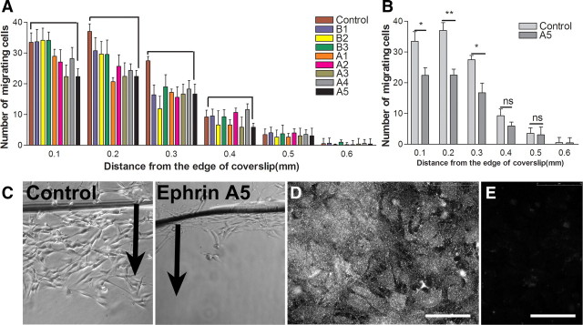
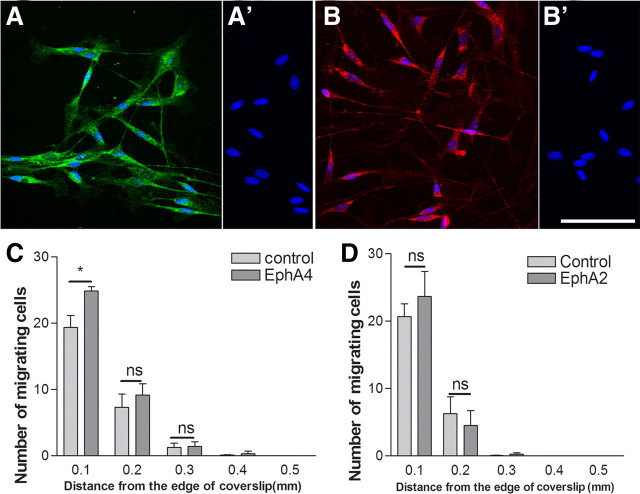
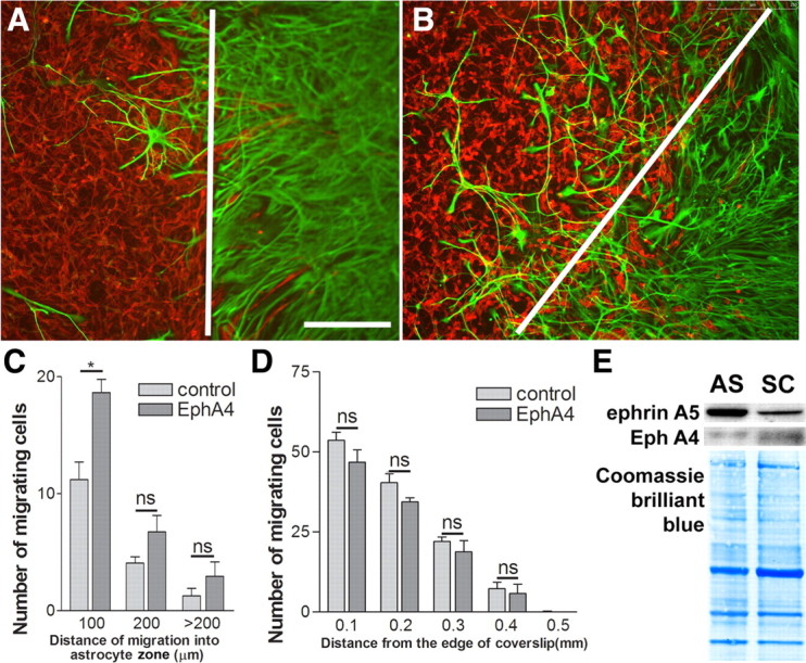
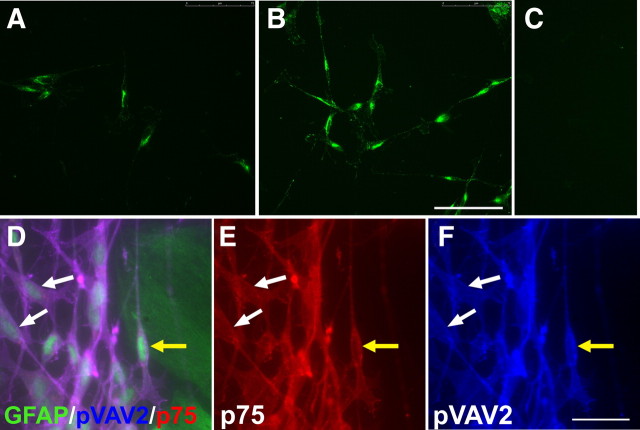
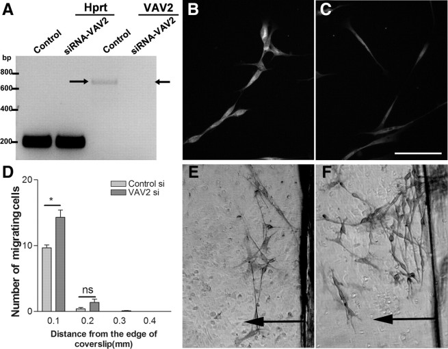
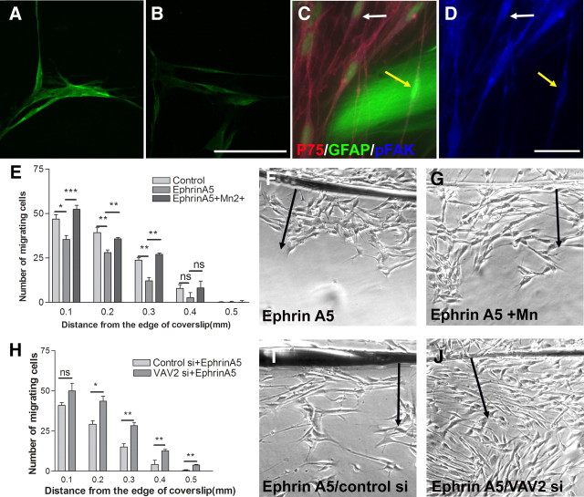
Schwann cells are a promising candidate for bridging spinal cord injuries and remyelinating axons. However, grafted Schwann cells show little intermingling with host astrocytes and therefore limited migration from transplant sites. This leads to the formation of a sharp border between host astrocytes and Schwann cells, which results in axons stalling at the graft-host interface and failing to exit the graft. We investigated the possibility that Eph/ephrin interactions are involved in the segregation of Schwann cells and astrocytes and in limiting Schwann cell migration. Using reverse transcription-PCR, we have characterized the ephrin and Eph profile in cultured Schwann cells and astrocytes, showing that astrocytes produce all the ephrinAs and Schwann cells produce the receptors EphA2, EphA4, and EphA7. Several ephrinAs inhibit Schwann cell migration on laminin, with ephrinA5 being the most effective. Blocking the EphA receptors with excess EphA4-Fc increases Schwann cell migration on astrocytes and improves Schwann-astrocyte intermingling. We show that the action of ephrinA5 on Schwann cells is mediated via VAV2. Both clustered ephrinA5 and astrocyte contact increases the phosphorylation of VAV2 in Schwann cells. Knockdown of VAV2 abrogates the inhibitory effect of clustered ephrinA5 on migration and increases the ability of Schwann cells to migrate on astrocytes. In addition, we found a role for ephrinA5 in inhibiting Schwann cell integrin signaling and function. Overall, we suggest that Eph/ephrin interactions inhibit Schwann cell migration and intermingling with astrocytes via VAV signaling affecting integrin function.


Figures

Figure 1.
Figure 1.
The effect of clustered ephrins on Schwann cell migration on laminin. A, EphrinA1, ephrinA3, and ephrinA5 reduce Schwann cell migration on laminin with the greatest reduction caused by ephrinA5. Clustered ephrinBs did not affect Schwann cell migration on laminin. B, Clustered ephrinA5 reduces the number of Schwann cells migrating from the edge of coverslip significantly. n = 3; t test, *p < 0.05. C, Images of Schwann cell migration on laminin in the control nontreated and ephrinA5-treated group. Arrows point to the direction of cell migration from the edge of coverslip. D, E, Immunostaining for ephrinA5 on astrocytic monolayers (D) and its corresponding negative control (E). Scale bars, 75 μm.
Figure 2.
Figure 2.
The effect of Eph–Fc in solution on Schwann cell migration on astrocytes. A–B′, Immunostaining for EphA4 (A) and EphA2 (B) in postnatal Schwann cells and their corresponding negative controls (A′, B′). DAPI nuclear staining is blue. Scale bar, 75 μm. C, D, EphA4–Fc improves Schwann cell migration on astrocytes near the coverslip in the Schwann cell migration assay, but EphA2–Fc did not have any effect. n = 3; t test, *p < 0.05.
Figure 3.
Figure 3.
The effect of EphA4–Fc in solution on Schwann cell intermingling with astrocytes in the boundary assay. A, B, Schwann cells labeled for p75 (red) and astrocytes for GFAP (green) in the boundary assay in control (A) and EphA4–Fc-treated (B) confrontation cultures. The white lines depicts the boundaries between the astrocyte cell front and Schwann cells. Scale bar, 250 μm. C, EphA4–Fc (10 μg/ml) increases Schwann cell–astrocyte intermingling in the boundary assay. n = 3; t test, *p < 0.05. D, Schwann cell migration on laminin is not affected by EphA4–Fc in solution, excluding reverse signaling through Schwann cell ephrins. n = 3. E, Immunoblotting of ephrinA5 and EphA4 on the membrane preparations from Schwann cells (SC) and astrocytes (AS). It demonstrates a higher level of ephrinA5 on astrocytes than Schwann cells. A reverse trend is seen for EphA4 that shows a higher level on Schwann cells than astrocytes. Coomassie Brilliant Blue staining on the blots demonstrates equal loading of proteins.
Figure 4.
Figure 4.
The effect of ephrinA5 on phosphorylation of VAV2 in Schwann cells. A–C, Immunostaining for phosphorylated VAV2 in nontreated control (A), EphrinA5-treated Schwann cells (B), and negative control (C). Scale bar, 75 μm. Addition of clustered ephrinA5 to Schwann cells plated on laminin leads to an increase in the levels of pVAV2 at 10 min. D–F, Immunostaining of Schwann cells labeled for p75 (red) in contact with GFAP-labeled (green) astrocytes (D). E and F show the corresponding p75 and phosphorylated VAV2 staining, respectively. The yellow arrow points to a Schwann cell in contact with astrocytes. There are also piled up Schwann cells in contact with astrocytes adjoining it. The white arrows point to noncontacting Schwann cells. Schwann cells in contact with astrocytes have increased levels of phosphorylated VAV2 compared with control noncontacting cells. Scale bar, 25 μm.
Figure 5.
Figure 5.
The effect of VAV2 knockdown on Schwann cell migration on astrocytes. A, Assessment of VAV2 knockdown at 48 h using RT-PCR. Note the complete absence of a 663 bp band (arrow) corresponding to VAV2 in the VAV2 siRNA-treated group only. B, C, VAV2 immunostaining in control (B) and VAV2 siRNA-treated cells (C). Assessment of VAV2 knockdown at 48 h by immunostaining shows protein levels of ∼50% of control at the time of start of the migration assays. D, Knockdown of VAV2 increases the number of Schwann cells migrating from the edge of coverslip compared with control cells. n = 3; t test, *p < 0.05. E, F, Schwann cell migration on astrocytic monolayers in the control siRNA-treated (E) and VAV2 siRNA-treated (F) Schwann cells. Arrows point to the direction of cell migration.
Figure 6.
Figure 6.
The effect of ephrins on integrin-mediated signaling and migration. A, B, pFAK immunostaining in nontreated controls (A) and clustered ephrinA5-treated cells (B). Addition of clustered ephrinA5 to Schwann cells plated on laminin reduces pFAK levels. Scale bar, 75 μm. C, D, Immunostaining of Schwann cells labeled for p75 in contact with GFAP-labeled astrocytes (C) and the corresponding phosphorylated FAK staining (D). Schwann cells in contact with hypertrophied astrocytes have decreased pFAK levels compared with control noncontacting Schwann cells. The yellow arrow points to a Schwann cell in contact with astrocytes and the white arrow to a noncontacting Schwann cell. Scale bar, 25 μm. E, Addition of integrin activating Mn2+ (500 μm) to Schwann cells migrating in the presence of clustered ephrinA5 reverses the inhibitory effect of ephrinA5 on migration. n = 5; ANOVA, *p < 0.05. F, G, Images of the migration assay after addition of clustered ephrinA5 in the absence (F) and presence (G) of Mn2+. H, VAV2 knockdown in Schwann cells abolishes the inhibitory effect of ephrinA5 on Schwann cell migration on laminin. n = 3; ANOVA, *p < 0.05. I, J, Images of Schwann cell migration on laminin in the presence of clustered ephrinA5 in control siRNA (I) and VAV2 siRNA (J) treated cells.

References

    1. Adcock KH, Brown DJ, Shearer MC, Shewan D, Schachner M, Smith GM, Geller HM, Fawcett JW. Axon behaviour at Schwann cell–astrocyte boundaries: manipulation of axon signalling pathways and the neural adhesion molecule L1 can enable axons to cross. Eur J Neurosci. 2004;20:1425–1435. - PubMed
    1. Andrews MR, Stelzner DJ. Evaluation of olfactory ensheathing and schwann cells after implantation into a dorsal injury of adult rat spinal cord. J Neurotrauma. 2007;24:1773–1792. - PubMed
    1. Biernaskie J, Sparling JS, Liu J, Shannon CP, Plemel JR, Xie Y, Miller FD, Tetzlaff W. Skin-derived precursors generate myelinating Schwann cells that promote remyelination and functional recovery after contusion spinal cord injury. J Neurosci. 2007;27:9545–9559. - PMC - PubMed
    1. Bourgin C, Murai KK, Richter M, Pasquale EB. The EphA4 receptor regulates dendritic spine remodeling by affecting beta1-integrin signaling pathways. J Cell Biol. 2007;178:1295–1307. - PMC - PubMed
    1. Brockes JP, Fields KL, Raff MC. Studies on cultured rat Schwann cells. I. Establishment of purified populations from cultures of peripheral nerve. Brain Res. 1979;165:105–118. - PubMed

Publication types

MeSH terms