The functional properties of barrel cortex neurons projecting to the primary motor cortex
- PMID: 20335461
- PMCID: PMC6634518
- DOI: 10.1523/JNEUROSCI.3774-09.2010
The functional properties of barrel cortex neurons projecting to the primary motor cortex
Abstract
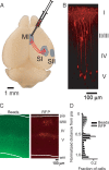
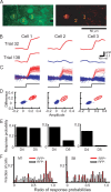
Nearby neurons, sharing the same locations within the mouse whisker map, can have dramatically distinct response properties. To understand the significance of this diversity, we studied the relationship between the responses of individual neurons and their projection targets in the mouse barrel cortex. Neurons projecting to primary motor cortex (MI) or secondary somatosensory area (SII) were labeled with red fluorescent protein (RFP) using retrograde viral infection. We used in vivo two-photon Ca(2+) imaging to map the responses of RFP-positive and neighboring L2/3 neurons to whisker deflections. Neurons projecting to MI displayed larger receptive fields compared with other neurons, including those projecting to SII. Our findings support the view that intermingled neurons in primary sensory areas send specific stimulus features to different parts of the brain.
Figures


Similar articles
-
Vibrissal motor cortex in the rat: connections with the barrel field.Exp Brain Res. 1995;104(1):41-54. doi: 10.1007/BF00229854. Exp Brain Res. 1995. PMID: 7621940
-
Whisker trimming begun at birth or on postnatal day 12 affects excitatory and inhibitory receptive fields of layer IV barrel neurons.J Neurophysiol. 2005 Dec;94(6):3987-95. doi: 10.1152/jn.00569.2005. Epub 2005 Aug 10. J Neurophysiol. 2005. PMID: 16093330
-
The functional microarchitecture of the mouse barrel cortex.PLoS Biol. 2007 Jul;5(7):e189. doi: 10.1371/journal.pbio.0050189. Epub 2007 Jul 10. PLoS Biol. 2007. PMID: 17622195 Free PMC article.
-
A Cellular Resolution Map of Barrel Cortex Activity during Tactile Behavior.Neuron. 2015 May 6;86(3):783-99. doi: 10.1016/j.neuron.2015.03.027. Epub 2015 Apr 23. Neuron. 2015. PMID: 25913859
-
Spatial organization of neuronal population responses in layer 2/3 of rat barrel cortex.J Neurosci. 2007 Nov 28;27(48):13316-28. doi: 10.1523/JNEUROSCI.2210-07.2007. J Neurosci. 2007. PMID: 18045926 Free PMC article.
Cited by
-
Structure of a single whisker representation in layer 2 of mouse somatosensory cortex.J Neurosci. 2015 Mar 4;35(9):3946-58. doi: 10.1523/JNEUROSCI.3887-14.2015. J Neurosci. 2015. PMID: 25740523 Free PMC article.
-
Highly differentiated projection-specific cortical subnetworks.J Neurosci. 2011 Jul 13;31(28):10380-91. doi: 10.1523/JNEUROSCI.0772-11.2011. J Neurosci. 2011. PMID: 21753015 Free PMC article.
-
Pathway-specific reorganization of projection neurons in somatosensory cortex during learning.Nat Neurosci. 2015 Aug;18(8):1101-8. doi: 10.1038/nn.4046. Epub 2015 Jun 22. Nat Neurosci. 2015. PMID: 26098757
-
Projection-specific Activity of Layer 2/3 Neurons Imaged in Mouse Primary Somatosensory Barrel Cortex During a Whisker Detection Task.Function (Oxf). 2020 Jul 2;1(1):zqaa008. doi: 10.1093/function/zqaa008. eCollection 2020. Function (Oxf). 2020. PMID: 35330741 Free PMC article.
-
Target dependence of orientation and direction selectivity of corticocortical projection neurons in the mouse V1.Front Neural Circuits. 2013 Sep 23;7:143. doi: 10.3389/fncir.2013.00143. eCollection 2013. Front Neural Circuits. 2013. PMID: 24068987 Free PMC article.
References
-
- Alloway KD. Information processing streams in rodent barrel cortex: The differential functions of barrel and septal circuits. Cereb Cortex. 2008;18:979–989. - PubMed
-
- Armstrong-James M, Fox K. Spatiotemporal convergence and divergence in the rat S1 “barrel” cortex. J Comp Neurol. 1987;263:265–281. - PubMed
MeSH terms
Substances
LinkOut - more resources
Full Text Sources
Miscellaneous
