. 2010 Mar 24;30(12):4273-84.
doi: 10.1523/JNEUROSCI.4017-09.2010.

TASK channels contribute to the K+-dominated leak current regulating respiratory rhythm generation in vitro

Affiliations

TASK channels contribute to the K+-dominated leak current regulating respiratory rhythm generation in vitro

Hidehiko Koizumi et al. J Neurosci. .

Abstract

Leak channels regulate neuronal activity and excitability. Determining which leak channels exist in neurons and how they control electrophysiological behavior is fundamental. Here we investigated TASK channels, members of the two-pore domain K(+) channel family, as a component of the K(+)-dominated leak conductance that controls and modulates rhythm generation at cellular and network levels in the mammalian pre-Bötzinger complex (pre-BötC), an excitatory network of neurons in the medulla critically involved in respiratory rhythmogenesis. By voltage-clamp analyses of pre-BötC neuronal current-voltage (I-V) relations in neonatal rat medullary slices in vitro, we demonstrated that pre-BötC inspiratory neurons have a weakly outward-rectifying total leak conductance with reversal potential that was depolarized by approximately 4 mV from the K(+) equilibrium potential, indicating that background K(+) channels are dominant contributors to leak. This K(+) channel component had I-V relations described by constant field theory, and the conductance was reduced by acid and was augmented by the volatile anesthetic halothane, which are all hallmarks of TASK. We established by single-cell RT-PCR that pre-BötC inspiratory neurons express TASK-1 and in some cases also TASK-3 mRNA. Furthermore, acid depolarized and augmented bursting frequency of pre-BötC inspiratory neurons with intrinsic bursting properties. Microinfusion of acidified solutions into the rhythmically active pre-BötC network increased network bursting frequency, halothane decreased bursting frequency, and acid reversed the depressant effects of halothane, consistent with modulation of network activity by TASK channels. We conclude that TASK-like channels play a major functional role in chemosensory modulation of respiratory rhythm generation in the pre-Bötzinger complex in vitro.


Figures

Figure 1.
Figure 1.
Overview of experimental in vitro neonatal rat rhythmic slice preparation with inspiratory neuronal imaging, set-up for local microinfusion in the pre-BötC, and representative current- and voltage-clamp recordings of functionally identified inspiratory neurons in vitro. A, B, Glass pipette suction electrodes on hypoglossal (XII) nerves (A) were used to bilaterally record inspiratory motoneuronal population discharge (B, integrated population activity ∫XII) as a monitor respiratory network rhythmic activity. Inspiratory neurons were localized for whole-cell recording in the pre-BötC by imaging single-neuron (box at lower left, ×63 magnification) and population fluorescence transients (pseudo-colored fluorescence flash images shown in A), produced by inspiratory activity of neurons retrogradely labeled with the Ca2+-sensitive dye Calcium Green-1 AM (see Materials and Methods for detailed explanation). Imaged population activity (×20 fluorescence population flash images shown superimposed bilaterally on IR-DIC image of ventral half of slice in A) was also used to locate the core of active pre-BötC inspiratory neurons to guide insertion of micropipettes into the center of the inspiratory population for local microinfusion of aCSF containing acid or halothane. C, Imaged pre-BötC inspiratory neurons were recorded in current-clamp and further identified functionally by rhythmic bursting in synchrony with XII motor discharge. Neurons with intrinsic voltage-dependent bursting properties (bottom traces in C) were initially identified and distinguished from nonintrinsic bursters by rhythmic ectopic bursting (marked by dots above bursts) unsynchronized with XII discharge when the neuronal membrane potential (Vm) was depolarized by steady current injection. D, I-V relations of identified inspiratory neurons were measured by a voltage-clamp ramp protocol (30 mV/s ramp) with synaptic transmission blocked under control conditions and after bath-application of acidified aCSF and/or aCSF containing halothane. I-V relation for a nonintrinsic burster neuron is shown. Leak I-V relations, used to calculate leak conductances, were determined from linear regression (solid gray line shown) to the passive regions (below −65 mV) of I-V curves. The reversal potential, ELeak, of the total leak current (0 current intercept of regression line), is indicated by the vertical dashed line and was typically depolarized from the calculated K+ equilibrium potential (EK indicated) for the recording conditions.
Figure 2.
Figure 2.
Acid decreases and halothane increases leak conductance of pre-BötC inspiratory neurons. A, B, Representative examples of whole-cell voltage-clamp ramp (30 mV/s) recordings of I-V relations of pre-BötC inspiratory neurons (data from nonintrinsic bursters in both panels) before (gray curves) and after (black) bath-application of acidic (pH = 6.8) aCSF (A) or halothane-containing aCSF (2%, B), showing rotations of I-V relations about intersection at a membrane potential (ELeakK) corresponding closely to EK, which defines the K+ component of the total leak current. Inset (box) in A shows expanded view of I-V rotation about ELeakK (indicated by vertical dashed lines in all panels). Leak conductance (gLeak) is indicated by slope of linear regression fits to the passive segment (−100 to −70 mV) of each I-V ramp (solid lines). In the examples shown, gLeak decreased by 1.0 nS or by 20% from control value with acid and increased by 3.5 nS (67% above control conductance) with halothane. C, Acid (pH 6.8) reverses the increase of gLeak by halothane (green I-V relation) in a representative intrinsically bursting inspiratory neuron. Coapplication of acid (pH 6.8) + halothane (2%) (red curve) restores gLeak to that of control I-V relation (black); red I-V relation is superimposed on black curve in the inset showing expanded view of I-V relations at voltages below and above ELeakK over which gLeak was measured. Blue curve shows I-V relation during exposure to acid alone.
Figure 3.
Figure 3.
TASK-like leak I-V relations are fit by the curve predicted from constant field theory for electrodiffusion through an open K+-selective pore. A, B, Representative ramp I-V relations of different inspiratory neurons (nonintrinsic burster in A, intrinsic burster in B) exposed to acidic aCSF (pH 7.0, A) and aCSF containing halothane (2%, B) when currents other than TASK are blocked. Nav+ channels were blocked with TTX (1 μm), Kir was blocked with Ba2+ (200 μm), Kv+ with extracellular TEA (10 mm), Ca2+ channels with Cd2+ (200 μm), and Ih blocked with ZD7288 (100 μm). Difference I-V curves (red = control-acid in A, red = halothane-control in B) have a shape similar to the curve (green) calculated by the Goldman-Hodgkin-Katz (GHK) constant field equation (see Materials and Methods) with values of intra- and extracellular K+ concentrations (125 mm and 8 mm, respectively) for the recording conditions. Difference (red) I-V relation in B obscures control curve (gray). Equilibrium potentials (0 current potentials, ELeakK, vertical dashed lines) of currents induced by acid and halothane are identical and near EK.
Figure 4.
Figure 4.
Pre-BötC inspiratory neurons contain TASK-1 and in some cases TASK-3 mRNA. Electrophoresis gel generated by scRT-PCR from mRNA in cytoplasm aspirated from this inspiratory neuron (intrinsic burster) shows TASK-1 (column 2) and TASK-3 (column 3) cDNA. The band for TASK-1 corresponds to the TASK-1 reference [column 6, positive control from 100 pg of total rat brain RNA (tRNA)] and to the predicted molecular weight of amplimers of TASK-1 (512 bp, column at left). The band for TASK-3 corresponds to the TASK-3 reference (column 7 positive control) and to the predicted molecular weight of amplimers of TASK-3 (763 bp). No TASK-1 or TASK-3 signals were present in negative controls (nCTR, columns 4 and 5 in this example) from ‘mock harvests.’
Figure 5.
Figure 5.
Modulation of intrinsic bursting frequency of pre-BötC inspiratory neurons by acid. A, Current-clamp recording illustrating that transiently applied acidic aCSF (pH 6.8) reversibly depolarizes (maximally by ∼7 mV in this example) and increases the baseline voltage-dependent rhythmic bursting frequency of a representative intrinsic burster synaptically isolated after blocking Ca2+ channels with Cd2+ (200 μm). B, Traces with expanded time scale show bursting patterns before and during (epochs indicated by solid bars and arrows) bath application of acidic aCSF.
Figure 6.
Figure 6.
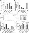
Modulation of respiratory network inspiratory bursting frequency in vitro by acid, halothane, and modulatory interactions of acid and halothane. A, B, Examples of progressive and reversible increase of inspiratory XII motoneuronal bursting frequency with acidic aCSF pH (A), and reversible decrease in bursting frequency with progressive increments of aCSF halothane concentrations (0.5% to 1.5%) (B) from two representative slices. Bars represent mean steady-state burst frequencies (bursts/min) averaged over 10 to 20 consecutive bursts at levels of aCSF pH or halothane concentration indicated. C, Example of rectified, integrated hypoglossal nerve inspiratory discharge (∫XII) showing elimination of rhythmic bursting by halothane and restoration of bursting by coapplied acid (pH 6.5) in the continued presence of halothane. D, Example of a full diagnostic sequence for involvement of TASK-like channels in control of network rhythm by acid and halothane. Acidic aCSF (pH 6.5) increased steady-state inspiratory burst frequency, halothane (0.5% after recovery from acid) decreased burst frequency, and acid (pH 6.5) dominated over halothane to increase burst frequency above control. E, Summary data of steady-state XII burst frequency (% control) during slice exposure to acid, halothane, and acid + halothane averaged over multiple slice experiments. Acid (pH 7.0 to 6.5, n = 11 slices pooled) increased mean inspiratory burst frequency, halothane (0.5% to 2.0%, n = 22 slices pooled) decreased burst frequency, and the effect of acid dominates over that of halothane when slices were exposed to both halothane and acid (n = 7 slices pooled). Bars are mean values and error bars are +SE.
Figure 7.
Figure 7.
Modulation of inspiratory rhythm by acid and halothane at the level of the pre-BötC network. A, Continuous microinfusion of acidic aCSF (pH 6.8, bar) bilaterally into the pre-BötC directly increased inspiratory burst frequency recorded from hypoglossal nerves (∫XII shown). Expansion of selected sections of A showing acid-induced increase of burst frequency, followed by return to control frequency, are shown at top. B, Inspiratory burst frequency versus time plot for A, continuously computed by taking the inverse of each interburst interval and smoothing with a three-point binomial filter. C, Data as in A pooled from five slices and four values of pH of aCSF separately microinfused into the pre-BötC in each slice. Network bursting frequency increased monotonically with progressive local acidification. Solid line is a Hill equation fit to the data points with a Hill slope of 0.84. Error bars represent ±SD. D, Example of burst frequency versus time plot as in B computed for this slice by averaging the inverse of every two interburst intervals. Halothane (2% in aCSF solution) microinfused into the pre-BötC nearly stopped network rhythmic activity, whereas acidic aCSF (pH 6.8) applied in the bath during halothane microinfusion reversed the depressant effect of halothane and elevated network-bursting frequency above control values. E, Sequence of halothane and acid application as in D summarizing pooled results from multiple slice experiments (n = 4). In all slices halothane (2%) microinfused into the pre-BötC progressively reduced, and in 3 slices stopped, network rhythmic activity, whereas acidic aCSF (pH 6.8) subsequently bath-applied during halothane microinfusion reversed the effect of halothane and elevated bursting frequency above control levels. Bars are mean values and error bars are +SE.
Figure 8.
Figure 8.
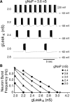
Bursting behavior of a model pre-BötC intrinsically bursting neuron modulated by changes in the K+ component (gLeakK) of the total leak conductance. A, Example simulation of rhythmic bursts of single neuron model when gNaP was set to 3.6 nS, near the upper end of the range of gNaP for pre-BötC intrinsic bursters (B). Traces are membrane potential versus time for simulated current-clamp recording conditions. Progressively increasing TASK-like gLeakK (from top to bottom simulation traces), corresponding to an increase in halothane concentration, progressively reduces neuronal burst frequency and ultimately stops rhythmic bursting (bottom). B, Summary plots of simulated relations between gLeakK and model neuron bursting frequency for different gNaP isopleth values covering the range of gNaP values (2.0 to 4.4 nS) found experimentally in pre-BötC intrinsically bursting inspiratory neurons (Purvis et al., 2007; Koizumi and Smith, 2008). The range of estimated gLeakK (2.0 to 7.0 nS) covers the measured experimental range of TASK-like K+ leak conductance with modulation by acid (lower end of range) and halothane (upper end). Model neuron burst frequency versus gLeakK over experimental range of gNaP shows that burst frequency is inversely proportional to gLeakK for all values of gNaP. The model neuron bursts rhythmically in the parameter range gLeakK = 2.0 to 3.0 nS and gNaP = 2.0 to 4.4 nS. At gLeakK below 2.0 nS, outside of the range of gLeakk estimated for acidic conditions in the present experiments, the neuron spikes tonically, whereas above gLeakK = 4.0 nS (halothane conditions) the neuron is silent over the range of gNaP values.
Figure 9.
Figure 9.
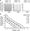
Modulation of rhythmic bursting behavior by population TASK-like K+ leak conductance in a model heterogeneous neuronal excitatory network of the pre-BötC. Simulation results shown are for a 50 excitatory neuron network model with a range of mean population K+ leak values (GleakK = 2.0 to 7.0 nS) covering the average range estimated from single neurons with acid and halothane modulation of GleakK. The simulated population had a range of GNaP (2.0 to 4.4 nS population mean values) as measured previously for a population of pre-BötC inspiratory neurons (Purvis et al., 2007; Koizumi et al., 2008) (for simplicity only simulation results for GNaP = 2.4 to 3.6 nS are shown). A, Example of model network simulation output when population GNaP was set for illustration to 2.6 nS (± 30%SD) and GLeakK was varied from 3.4 to 6.0 nS (± 30% SD). Shown are population-spiking activity histograms (50 ms bins) averaged over the 50 individual network neurons. Population burst frequency decreases as GLeakK increases, ultimately stopping network rhythmic activity, mimicking effects at the network level of halothane on TASK-like leak conductance. B, Summary plots of model relations between GLeakK and population bursting frequency for a range of fixed values of GNaP. For any value of GNaP, population-bursting frequency was inversely proportional to GLeakK, and network activity could be stopped at higher values of GLeakK. Below GNaP = 2.2 nS, the model network was silent. For GNaP = 2.4 to 3.6 nS, the network model exhibited rhythmic population bursting when GLeakK was between 2.4 to 4.8 nS (for simplicity only simulation results with GLeakK values above 3.0 nS are shown). For estimated GLeakK during exposure of the slice or pre-BötC to halothane (6.0 to 7.0 nS), the model network was silent, reproducing experimental results. See text for further explanation.

Similar articles

Cited by

References

    1. Bayliss DA, Siros JE, Talley EM. The TASK family: two-pore-domain background K+ channels. Mol Interven. 2003;3:205–219. - PubMed
    1. Berg AP, Talley EM, Manger JP, Bayliss DA. Motoneurons express heteromeric TWIK-related acid-sensitive K+ (TASK) channels containing TASK-1 (KCNK3) and TASK-3 (KCNK9) subunits. J Neurosci. 2004;24:6693–6702. - PMC - PubMed
    1. Burdakov D, Jensen LT, Alexopoulos H, Williams RH, Fearon IM, O'Kelly I, Gerasimenko O, Fugger L, Verkhratsky A. Tandem-pore K+ channels mediate inhibition of orexin neurons by glucose. Neuron. 2006;50:711–722. - PubMed
    1. Butera RJ, Jr, Rinzel J, Smith JC. Models of respiratory rhythm generation in the pre-Botzinger complex. I. Bursting pacemaker neurons. J Neurophysiol. 1999a;82:382–397. - PubMed
    1. Butera RJ, Jr, Rinzel J, Smith JC. Models of respiratory rhythm generation in the pre-Botzinger complex. II. Populations of coupled pacemaker neurons. J Neurophysiol. 1999b;82:398–415. - PubMed

Publication types

MeSH terms