. 2010 Mar 25;464(7288):549-53.
doi: 10.1038/nature08873.

Coronary arteries form by developmental reprogramming of venous cells

Affiliations

Coronary arteries form by developmental reprogramming of venous cells

Kristy Red-Horse et al. Nature. .

Abstract

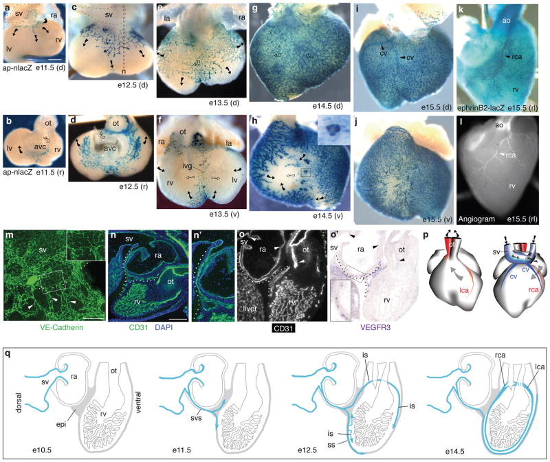
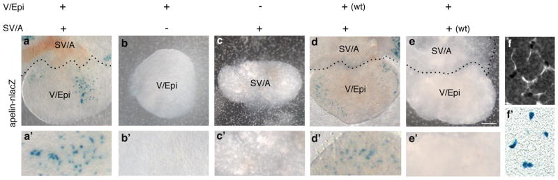
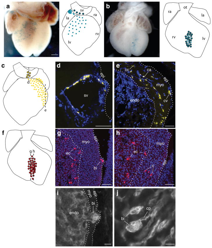
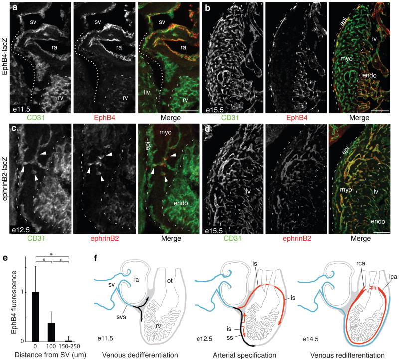
Coronary artery disease is the leading cause of death worldwide. Determining the coronary artery developmental program could aid understanding of the disease and lead to new treatments, but many aspects of the process, including their developmental origin, remain obscure. Here we show, using histological and clonal analysis in mice and cardiac organ culture, that coronary vessels arise from angiogenic sprouts of the sinus venosus-the vein that returns blood to the embryonic heart. Sprouting venous endothelial cells dedifferentiate as they migrate over and invade the myocardium. Invading cells differentiate into arteries and capillaries; cells on the surface redifferentiate into veins. These results show that some differentiated venous cells retain developmental plasticity, and indicate that position-specific cardiac signals trigger their dedifferentiation and conversion into coronary arteries, capillaries and veins. Understanding this new reprogramming process and identifying the endogenous signals should suggest more natural ways of engineering coronary bypass grafts and revascularizing the heart.


Figures

Figure 1
Figure 1. Coronary vessels sprout from the sinus venosus
a–j. X-gal stained apelin-nlacZ hearts from embryonic days indicated, shown in dorsal (d), rostral (r), and ventral (v) views. Coronary vessels (blue nuclei) originate near the sinus venosus (sv, outlined in a,c) and spread dorsally (a, c, e, g) and around the atrioventricular canal (avc) (b, d) and outflow tract (ot) (d) to the ventral interventricular groove (ivg) (f). Arrows indicate direction of new growth on the surface (filled) or in deeper layers (open). Inset in h is close up of boxed blood island-like structure. la, left atrium; ra, right atrium; rv, right ventricle; lv, left ventricle. Scale bar (for a–l), 200 μm. k. X-gal-stained e15.5 ephrinB2-lacZ heart (right lateral (rl) view) showing right coronary artery (rca, arrowhead). ao, aorta. l. Angiogram of e15.5 heart showing right coronary artery (rca, arrowhead). m. Frontal section through e11.5 heart immunostained for VE-cadherin. Coronary sprouts (green, arrowheads) are continuous with the SV (above dashed line). Inset is close up of boxed region showing junction between SV and coronary sprouts. Scale bar, 100 μm. n. Sagittal section (approximate section plane shown in c) through e12.5 heart stained for CD31 (green) and with nuclear dye DAPI (blue). Coronary sprout (dotted line) extends from the sv and courses around right atrium (ra) to reach right ventricle (rv). n’. Close-up of coronary sprout in n. o,o’. Adjacent sections from e11.5 heart immunostained for CD31 (o) and probed for VEGFR3 mRNA (purple) (o’). Coronary vessels (dotted line) express VEGFR3, an angiogenic sprout marker, whereas vessels formed by vasculogenesis (sv, atria, outflow tract) do not (arrowheads). Inset in o’ is close-up of boxed region. Scale bar (for n,o,o’), 200 μm. p. Schematic of right (rca) and left (lca) coronary arteries and coronary veins (cv) at e15.5. Arrows show blood flow direction. q. Schematic sagittal sections showing coronary sprout (blue) progression during development. epi, epicardium; svs, sinus venosus sprouts; ss, superficial sprouts; is, invading sprouts.
Figure 2
Figure 2. Analysis of coronary vessel sprouting in vitro
a–e. Intact heart (a) or dissected sinus venosus/atria (SV/A) or ventricle/epicardium (V/Epi) as indicated from e10.5 apelin-nlacZ embryos (or wild type (wt) where noted) cultured for 3 days at 37°C and stained with X-gal (blue nuclei) to show coronary development. Coronary sprouts formed only on intact hearts (a) or when dissected SV/A were recombined with V/Epi (d). Vessels arise from SV/A because no X-gal+ sprouts were detected when SV/A was from wild type (non-transgenic) embryo (e). a’–e’ Close up views. f,f’. Close-up of a cultured intact heart as in a stained for CD31 (f) and with X-gal (f’), confirming lacZ-positive (blue) nuclei are in coronary vessels. Scale bar, 200 μm.
Figure 3
Figure 3. Clonal analysis of coronary artery development
a–h. Micrographs and schematics of VE-cadherin-CreER-induced coronary clones at e13.5 (b,f) and e14.5 (a,c) marked with Rosa-lacZ (a,b) or Multi-color (ch), in which marked cells permanently express one of three different fluorescent reporters. Colored dots in schematics represent 10 cells in clone. Outlined dots are sister cells in SV or endocardium. a. Coronary clone (no. 24 in Table S1) with sister cells in sinus venosus (sv). Scale bar (for a,b), 200 μm. b. Coronary clone (no. 34) with sister cells in ventricular endocardium. c–e. Schematic (c) and two sections (d,e, dashed lines in c) of a coronary clone (no. 25) with sister cells in sinus venosus (d) and coronary artery (ca), coronary capillary (cc), and coronary vein (cv) (e). CD31 immunostaining (blue) shows all endothelial and endocardial cells. f–h. Schematic (f) and sections (g,h) of coronary clone (no. 28) with sister cells in coronary vessels (c), endocardial cells (ec), and adjacent blood island (bi). Nuclei are DAPI-stained (blue). Scale bars (d,e,g,f), 100 μm. i, j. Sections of e11.5 (i) and e12.5 (j) hearts immunostained for CD31 showing blood island-like structures budding from endocardium (endo) into myocardium (myo) (i) and joining the coronary vascular plexus (cp) (j). Scale bars, 25 μm. epi, epicardium; la, left atria; lv, left ventricle; ot, outflow tract; ra, right atria; rv, right ventricle.
Figure 4
Figure 4. Downregulation of venous markers and induction of arterial markers during coronary artery development
a. Sagittal section through heart of e11.5 EphB4-lacZ embryo immunostained for CD31 (green) and β-gal (EphB4, red). Coronary sprouts (dotted line) downregulate EphB4 as they migrate away from sinus venosus (sv). ra, right atria; rv, right ventricle; endo, endocardium; myo, myocardium; liv, liver. Scale bar, 100 μm. b. e15.5 EphB4-lacZ heart. Vessels on surface (epicardium, epi) have re-acquired EphB4 expression. Scale bar, 200 μm. c. Sagittal section through e12.5 ephrinB2-lacZ heart. Coronary sprouts that have invaded myocardium (green, arrowheads) upregulate artery/capillary marker ephrinB2 (red), whereas surface vessels (green, dashed line at left) do not. d. e15.5 ephrinB2-lacZ heart. EphrinB2 is expressed by all arteries and capillaries within myocardium but not by surface vessels. Scale bar, 100 μm. e. Quantification of EphB4 expression in coronary sprouts budding from sinus venosus (sv) at e11.5. Values are from double-stained hearts as in (a), normalized to CD31 staining at same position. Error bars, s.d. *, p<0.002 by Students t-test. f. Schematic showing changes in venous (blue) and arterial (red) marker expression during coronary development; black indicates dedifferentiated venous cells. svs, sinus venosus sprouts; ss, superficial sprouts; is, invading sprouts.

Comment in

Similar articles

Cited by

References

    1. Folkman J, Haudenschild C. Angiogenesis in vitro. Nature. 1980;288:551–556. - PubMed
    1. Leung DW, Cachianes G, Kuang WJ, Goeddel DV, Ferrara N. Vascular endothelial growth factor is a secreted angiogenic mitogen. Science. 1989;246:1306–1309. - PubMed
    1. Carmeliet P. Angiogenesis in life, disease and medicine. Nature. 2005;438:932–936. - PubMed
    1. Adams RH, Alitalo K. Molecular regulation of angiogenesis and lymphangiogenesis. Nat Rev Mol Cell Biol. 2007;8:464–478. - PubMed
    1. Ferrara N, Kerbel RS. Angiogenesis as a therapeutic target. Nature. 2005;438:967–974. - PubMed

Publication types