

Primary contribution to zebrafish heart regeneration by gata4(+) cardiomyocytes
- PMID: 20336144
- PMCID: PMC3040215
- DOI: 10.1038/nature08804
Primary contribution to zebrafish heart regeneration by gata4(+) cardiomyocytes
Abstract
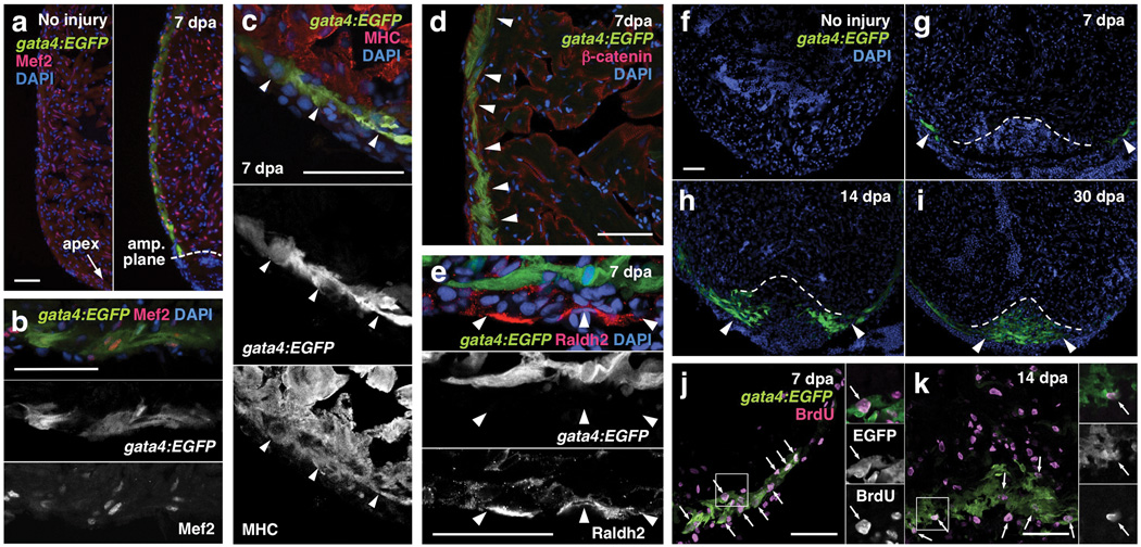
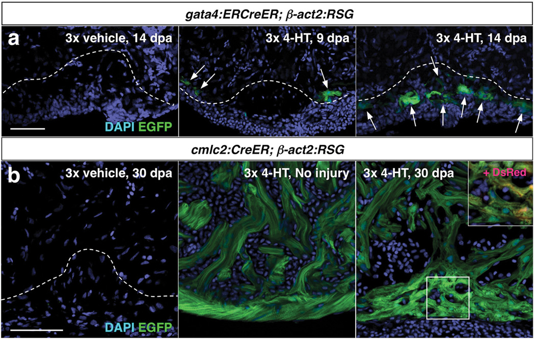
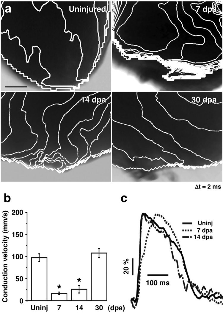
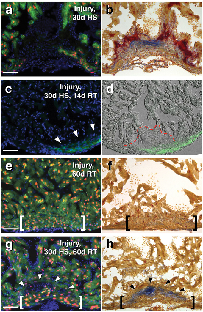
Recent studies indicate that mammals, including humans, maintain some capacity to renew cardiomyocytes throughout postnatal life. Yet, there is little or no significant cardiac muscle regeneration after an injury such as acute myocardial infarction. By contrast, zebrafish efficiently regenerate lost cardiac muscle, providing a model for understanding how natural heart regeneration may be blocked or enhanced. In the absence of lineage-tracing technology applicable to adult zebrafish, the cellular origins of newly regenerated cardiac muscle have remained unclear. Using new genetic fate-mapping approaches, here we identify a population of cardiomyocytes that become activated after resection of the ventricular apex and contribute prominently to cardiac muscle regeneration. Through the use of a transgenic reporter strain, we found that cardiomyocytes throughout the subepicardial ventricular layer trigger expression of the embryonic cardiogenesis gene gata4 within a week of trauma, before expression localizes to proliferating cardiomyocytes surrounding and within the injury site. Cre-recombinase-based lineage-tracing of cells expressing gata4 before evident regeneration, or of cells expressing the contractile gene cmlc2 before injury, each labelled most cardiac muscle in the ensuing regenerate. By optical voltage mapping of surface myocardium in whole ventricles, we found that electrical conduction is re-established between existing and regenerated cardiomyocytes between 2 and 4 weeks post-injury. After injury and prolonged fibroblast growth factor receptor inhibition to arrest cardiac regeneration and enable scar formation, experimental release of the signalling block led to gata4 expression and morphological improvement of the injured ventricular wall without loss of scar tissue. Our results indicate that electrically coupled cardiac muscle regenerates after resection injury, primarily through activation and expansion of cardiomyocyte populations. These findings have implications for promoting regeneration of the injured human heart.
Figures
Comment in
-
Thanks be to zebrafish.Circ Res. 2010 Sep 3;107(5):570-2. doi: 10.1161/RES.0b013e3181f6c515. Circ Res. 2010. PMID: 20814025 No abstract available.
References
-
- Quaini F, et al. Chimerism of the transplanted heart. N. Engl. J. Med. 2002;346(1):5–15. - PubMed
-
- Rubart M, Field LJ. Cardiac regeneration: Repopulating the heart. Annu. Rev. Physiol. 2006;68:29–49. - PubMed
-
- Poss KD, Wilson LG, Keating MT. Heart regeneration in zebrafish. Science. 2002;298(5601):2188–2190. - PubMed
Publication types
MeSH terms
Substances
Grants and funding
- HL007101/HL/NHLBI NIH HHS/United States
- HL007208/HL/NHLBI NIH HHS/United States
- HL081674/HL/NHLBI NIH HHS/United States
- HL054737/HL/NHLBI NIH HHS/United States
- R01 HL064282/HL/NHLBI NIH HHS/United States
- GM075846/GM/NIGMS NIH HHS/United States
- T32 HL007208/HL/NHLBI NIH HHS/United States
- HL064282/HL/NHLBI NIH HHS/United States
- R01 HL081674/HL/NHLBI NIH HHS/United States
- R01 HL054737/HL/NHLBI NIH HHS/United States
- R21 GM075946/GM/NIGMS NIH HHS/United States
- T32 HL007101/HL/NHLBI NIH HHS/United States
- R01 HL109264/HL/NHLBI NIH HHS/United States
- HHMI/Howard Hughes Medical Institute/United States
LinkOut - more resources
Full Text Sources
Other Literature Sources
Molecular Biology Databases
Research Materials
Miscellaneous
