. 2010 Mar 25;464(7288):606-9.
doi: 10.1038/nature08899.

Zebrafish heart regeneration occurs by cardiomyocyte dedifferentiation and proliferation

Affiliations

Zebrafish heart regeneration occurs by cardiomyocyte dedifferentiation and proliferation

Chris Jopling et al. Nature. .

Abstract

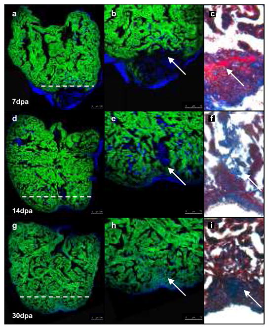
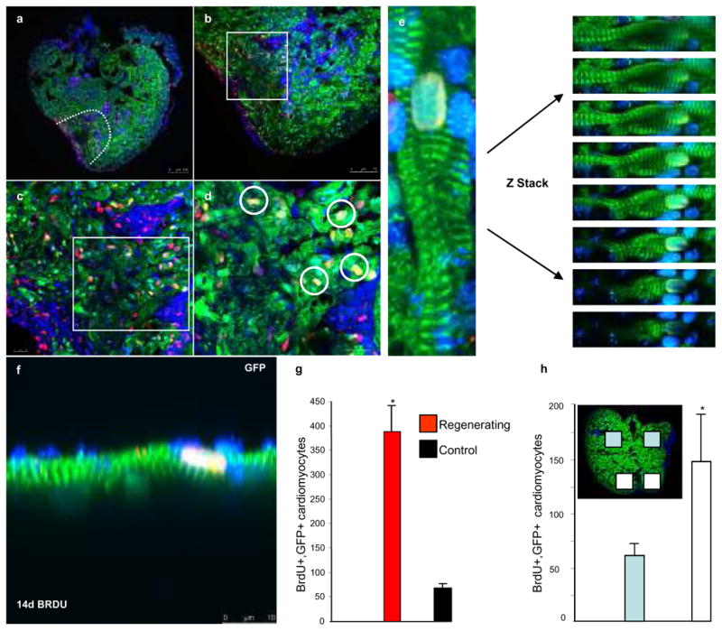
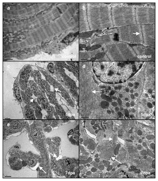
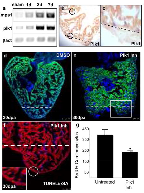
Although mammalian hearts show almost no ability to regenerate, there is a growing initiative to determine whether existing cardiomyocytes or progenitor cells can be coaxed into eliciting a regenerative response. In contrast to mammals, several non-mammalian vertebrate species are able to regenerate their hearts, including the zebrafish, which can fully regenerate its heart after amputation of up to 20% of the ventricle. To address directly the source of newly formed cardiomyocytes during zebrafish heart regeneration, we first established a genetic strategy to trace the lineage of cardiomyocytes in the adult fish, on the basis of the Cre/lox system widely used in the mouse. Here we use this system to show that regenerated heart muscle cells are derived from the proliferation of differentiated cardiomyocytes. Furthermore, we show that proliferating cardiomyocytes undergo limited dedifferentiation characterized by the disassembly of their sarcomeric structure, detachment from one another and the expression of regulators of cell-cycle progression. Specifically, we show that the gene product of polo-like kinase 1 (plk1) is an essential component of cardiomyocyte proliferation during heart regeneration. Our data provide the first direct evidence for the source of proliferating cardiomyocytes during zebrafish heart regeneration and indicate that stem or progenitor cells are not significantly involved in this process.


Figures

Fig. 1
Fig. 1. Regenerated cardiomyocytes are derived from differentiated cardiomyocytes
Cardiomyocytes in transgenic zebrafish (tg-cmlc2a-Cre-Ert2: tg-cmlc2a-LnL-GFP) were genetically labelled at 48 hpf by inducing Cre activity with tamoxifen. These embryos were then grown to adulthood (3months/sexually mature) at which point the heart was amputated and allowed to regenerate for 7 (a, b, c), 14 (d, e, f) or 30 days (g, h, i). Dashed white line represents the plane of amputation. At 7 dpa (a, b) relatively little regeneration has occurred. Trichromic staining indicates a fibrin clot has formed adjacent to the wound (c). By 14 dpa, GFPpos cardiomyocytes have regenerated a substantial amount of new cardiac tissue (d, e) and the fibrin clot is reduced in size (f). At 30 dpa, heart regeneration is virtually complete (g, h) and all of the regenerated tissue is comprised of GFPpos cardiomyocytes. The clot has been replaced by a small scar (h). Scale bars represent 100 μm in a, d, g, and 75 μm in b, e, h.
Fig. 2
Fig. 2. Differentiated cardiomyocytes re-enter the cell cycle
Transgenic zebrafish (tg-cmlc2a-Cre-Ert2: tg-cmlc2a-LnL-GFP) genetically labelled at 48 hpf and grown to adulthood were amputated then treated with BrdU at 7 dpa, hearts were then isolated and processed at 14 dpa (a–f). Green= GFPpos cardiomyocytes; Red=BrdUpos cells; Blue= DAPI; Yellow= BrdUpos/GFPpos cardiomyocytes (white rings in d).(g) Indicates the average number of BrdUpos/GFPpos cardiomyocytes/section +/− SEM, t-test * p<0.01; amputated n=17 sections from 7 different animals, control n=9 sections from 3 different animals. (h and inset) Indicates the distribution of BrdUpos/GFPpos cardiomyocytes (n=5 sections from 5 different animals). Scale bars represent 100 μm in a, 75 μm in b, and 10 μm in c, d, f.
Fig. 3
Fig. 3. Cardiomyocytes dedifferentiate resulting in the disassembly of sarcomeric structure and detachment
Electron microscopy of a control heart (a, b), a 5-dpa regenerating heart (c, d) and a 7-dpa regenerating heart (e, f). Cardiomyocytes in unamputated control samples show a tightly organised sarcomeric structure (a), at higher magnification (b) the Z-lines are clearly visible (white arrow). At 5 dpa many of the cardiomyocytes display a disorganised sarcomeric structure (c) along with the appearance of intercellular spaces (white arrows). Closer examination reveals a loss of Z-lines (d, white arrow). At 7 dpa there is a similar loss of structure and appearance of intercellular spaces (e white arrows). At higher magnification (f) myosin fibres are visible (arrows) however both longitudinal (upper arrow) and transverse (lower arrow) fibres are present within the same cardiomyocyte indicating disorganised sarcomeric structure. Scale bars represent 0.5 μm in a, b, d, and 2 μm in c, e, f.
Fig. 4
Fig. 4. plk1 is necessary for cardiac regeneration
(a) Semi-quantitative RT-PCR of mps1 and plk1. (b, c) Double in-situ hybridization detecting plk1 mRNA (blue) and cmlc2a mRNA (red/brown) in a 7-dpa ventricle. The plane of amputation is indicated with a dashed line. (d) 30-dpa regenerating heart (DMSO treated). The plane of amputation is indicated with a dashed line. (e) 30-dpa regenerating heart (cyclapolin9 treated). The plane of amputation is indicated with a dashed line the white box indicates the area shown in (f). (f) Consecutive section taken from the same heart pictured in (e), labelled with TUNEL (green) and anti-α sarcomeric actin (red). The plane of amputation is indicated with a dashed line. Inset shows a TUNEL positive cardiomyocyte. (g) Plk1 inhibiton reduces the number of cardiomyocytes entering the cell cycle during heart regeneration. The graph indicates the average number of BrdUpos/GFPpos cardiomyocytes/section +/− SEM, t-test * p<0.01; Plk1 inhibitor (cyclapolin9) treated (n=30 sections from 10 different animals), untreated control (n=9 sections from 3 different animals). Scale bars represent 100 μm in d, e, and 50 μm in f.

Comment in

  • Thanks be to zebrafish.
    Williams R. Williams R. Circ Res. 2010 Sep 3;107(5):570-2. doi: 10.1161/RES.0b013e3181f6c515. Circ Res. 2010. PMID: 20814025 No abstract available.

Similar articles

Cited by

References

    1. Oberpriller JO, Oberpriller JC. Response of the adult newt ventricle to injury. J Exp Zool. 1974;187:249–253. - PubMed
    1. Neff AW, Dent AE, Armstrong JB. Heart development and regeneration in urodeles. Int J Dev Biol. 1996;40:719–725. - PubMed
    1. Flink IL. Cell cycle reentry of ventricular and atrial cardiomyocytes and cells within the epicardium following amputation of the ventricular apex in the axolotl, Amblystoma mexicanum: confocal microscopic immunofluorescent image analysis of bromodeoxyuridine-labeled nuclei. Anat Embryol (Berl) 2002;205:235–244. - PubMed
    1. Poss KD, Wilson LG, Keating MT. Heart regeneration in zebrafish. Science. 2002;298:2188–2190. - PubMed
    1. Raya A, et al. Activation of Notch signaling pathway precedes heart regeneration in zebrafish. Proc Natl Acad Sci U S A. 2003;100 (Suppl 1):11889–11895. - PMC - PubMed

Publication types

Substances