. 2010 Mar 9:1-5.
doi: 10.3928/15428877-20100215-30. Online ahead of print.

Optical Coherence Tomography Findings of Exophytic Retinal Capillary Hemangiomas of the Posterior Pole

Optical Coherence Tomography Findings of Exophytic Retinal Capillary Hemangiomas of the Posterior Pole

Eric K Chin et al. Ophthalmic Surg Lasers Imaging. .

Abstract

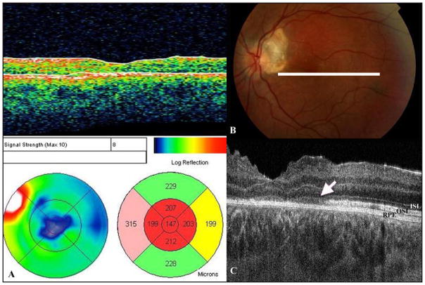
Exophytic retinal capillary hemangiomas (RCH) can be a diagnostic challenge in subjects without von Hippel-Lindau disease (VHL). This report of two cases describes the optical coherence tomographic (OCT) characteristics of RCH in two eyes of a subject with VHL and in one eye of an otherwise normal subject. Three different OCT instruments were used (Stratus, Cirrus and/or custom high resolution Fourier-domain OCT with 4.5 mum axial resolution) depending on availability. All instruments localized the tumor to the outer retina. A sharp border between the tumor and overlying inner retina was noted. The tumor bulged into the subretinal space and showed marked shadowing. Associated cystoid macular edema and subretinal fluid were noted. High-resolution Fourier-domain OCT showed a focal photoreceptor layer rip in the adjacent tumor-free macula in one eye with poor vision after treatment. OCT may be a useful tool in diagnosing RCH and studying associated morphologic changes.


Conflict of interest statement

The authors have no financial or proprietary interest in the materials presented herein.

Figures

Figure 1
Figure 1
(A) Fundus photograph OD of Case #1 showing a sub-retinal mass and a circinate ring of exudates extending to the fovea. (B) Fundus photograph OS of Case #1 showing a peripapillary submacular mass. (C) and (D) Fluorescein angiography of OD and OS, respectively, showing intense hyperfluorescence of the mass with minimal leakage. (E) Cirrus Fourier-domain optical coherence tomography (Fd-OCT) image of the subretinal mass OD showing a lesion contiguous with the outer retina with dense shadowing with adjacent cystoid macular edema. (F) Cirrus Fd-OCT image of subretinal mass OS showing a lesion contiguous with the outer macula with dense shadowing. Adjacent cystoid macular edema and subretinal fluid are also seen.
Figure 2
Figure 2
(A) Fundus photography OD of Case # 2 showing a peripapillary subretinal mass with associated retinal striae and hemorrhages, and serous macular detachment. Some peripapillary subretinal gliosis is seen over the mass. (B) Fluorescein angiography showing intense late view hyperfluorescence of the mass with minimal leakage. (C) Stratus Optical Coherence Tomography (OCT) image OS showing the mass contiguous with the outer retina with associated macular detachment. The macula adjacent to the mass (*) shows loss of the outer retinal layers. (D) High resolution Fourier-domain OCT image OS showing a peripapillary round mass bulging from the outer retina into the subretinal space with intense shadowing. (E) High resolution Fourier-domain OCT image of the left macula (*) adjacent to the mass showing severe disruption of the photoreceptor layer which appears partially ripped from the overlying detached macula (arrow). The high resolution Fourier-domain OCT instrument used was developed at the University of California Davis and provides axial resolution of 4.5 μm and transverse resolution 10 to 15 μm. ON—optic nerve; RCH—retinal capillary hemangioma
Figure 3
Figure 3
(A) Stratus Optical Coherence Tomography (OCT) image OS of Case #2 4 weeks after thermal laser photocoagulation of the retinal hemangioma showing complete resolution of the macular detachment and some thinning of the macula. (B) Fundus photography OS of Case #2 after laser treatment. The white line represents the orientation of the scan in Fig 3C. (C) High resolution Fourier-domain OCT image OS of Case # 2 6 months after laser treatment showing diffuse loss of the outer segment layer photoreceptors in the central macula (arrow). ISL-- inner segment layer; OSL-- outer segment layer; RPE-- retinal pigmented epithelium

References

    1. Gass JDM. Steroscopic Atlas of Macular Diseases Diagnosis and Treatment. St. Louis, Mo: Mosby-Year Book, Inc; 1997. Retinal Vascular Hamartomas; pp. 850–858.
    1. Alam S, Zawadzki RJ, Choi SS, et al. Clinical applications of rapid serial Fourier-domain optical coherence tomography for macular imaging. Ophthalmology. 2006;113:1425–1431. - PMC - PubMed
    1. Kreusel KM, Bechrakis NE, Neumann HPH, et al. Solitary juxtapapillary capillary retinal angioma and von Hippel-Lindau disease. Can J Ophthalmol. 2007;42:251–255. - PubMed
    1. McDonald HR. Diagnostic and therapeutic challenges. Retina. 2003;23:86–91. - PubMed
    1. Shields CL, Benevides R, Materin MA, Shields JA. Optical coherence tomography of retinal astrocytic hamartoma in 15 cases. Ophthalmology. 2006;113:1553–1557. - PubMed

LinkOut - more resources