

Neuromuscular junction formation between human stem-cell-derived motoneurons and rat skeletal muscle in a defined system
- PMID: 20337513
- PMCID: PMC2988647
- DOI: 10.1089/ten.TEC.2010.0040
Neuromuscular junction formation between human stem-cell-derived motoneurons and rat skeletal muscle in a defined system
Abstract
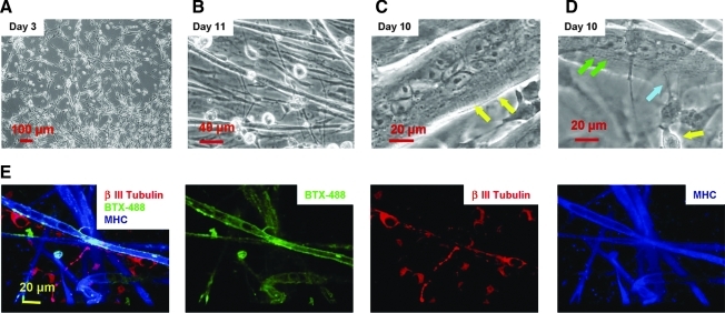
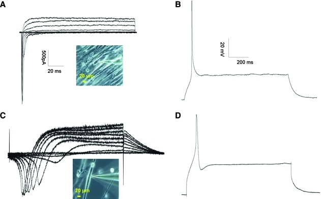
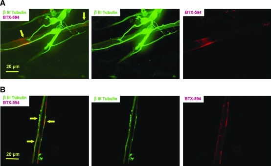
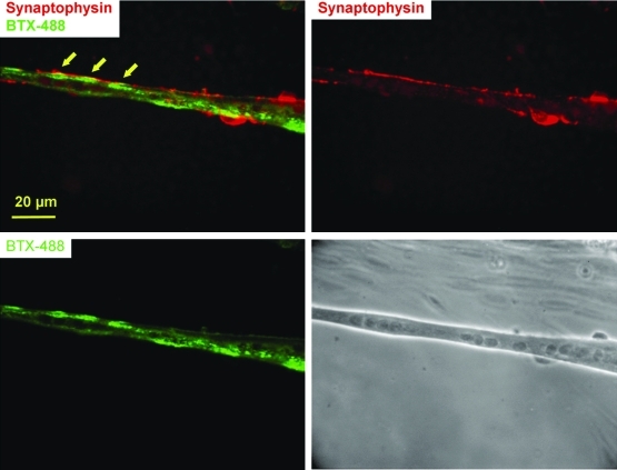
To date, the coculture of motoneurons (MNs) and skeletal muscle in a defined in vitro system has only been described in one study and that was between rat MNs and rat skeletal muscle. No in vitro studies have demonstrated human MN to rat muscle synapse formation, although numerous studies have attempted to implant human stem cells into rat models to determine if they could be of therapeutic use in disease or spinal injury models, although with little evidence of neuromuscular junction (NMJ) formation. In this report, MNs differentiated from human spinal cord stem cells, together with rat skeletal myotubes, were used to build a coculture system to demonstrate that NMJ formation between human MNs and rat skeletal muscles is possible. The culture was characterized by morphology, immunocytochemistry, and electrophysiology, while NMJ formation was demonstrated by immunocytochemistry and videography. This defined system provides a highly controlled reproducible model for studying the formation, regulation, maintenance, and repair of NMJs. The in vitro coculture system developed here will be an important model system to study NMJ development, the physiological and functional mechanism of synaptic transmission, and NMJ- or synapse-related disorders such as amyotrophic lateral sclerosis, as well as for drug screening and therapy design.
Figures
Similar articles
-
Neuromuscular junction formation between human stem cell-derived motoneurons and human skeletal muscle in a defined system.Biomaterials. 2011 Dec;32(36):9602-11. doi: 10.1016/j.biomaterials.2011.09.014. Epub 2011 Sep 23. Biomaterials. 2011. PMID: 21944471 Free PMC article.
-
Differentiation of glial cells and motor neurons during the formation of neuromuscular junctions in cocultures of rat spinal cord explant and human muscle.J Comp Neurol. 2001 Sep 17;438(2):239-51. doi: 10.1002/cne.1312. J Comp Neurol. 2001. PMID: 11536191
-
Origin of acetylcholinesterase in the neuromuscular junction formed in the in vitro innervated human muscle.Eur J Neurosci. 2004 Dec;20(11):2865-71. doi: 10.1111/j.1460-9568.2004.03752.x. Eur J Neurosci. 2004. PMID: 15579140
-
Optimization of Application-Driven Development of In Vitro Neuromuscular Junction Models.Tissue Eng Part B Rev. 2022 Dec;28(6):1180-1191. doi: 10.1089/ten.TEB.2021.0204. Epub 2022 Aug 1. Tissue Eng Part B Rev. 2022. PMID: 35018825 Free PMC article. Review.
-
How to Build and to Protect the Neuromuscular Junction: The Role of the Glial Cell Line-Derived Neurotrophic Factor.Int J Mol Sci. 2020 Dec 24;22(1):136. doi: 10.3390/ijms22010136. Int J Mol Sci. 2020. PMID: 33374485 Free PMC article. Review.
Cited by
-
Bioreactor model of neuromuscular junction with electrical stimulation for pharmacological potency testing.Integr Biol (Camb). 2017 Dec 11;9(12):956-967. doi: 10.1039/c7ib00144d. Integr Biol (Camb). 2017. PMID: 29168874 Free PMC article.
-
Tissue engineering the monosynaptic circuit of the stretch reflex arc with co-culture of embryonic motoneurons and proprioceptive sensory neurons.Biomaterials. 2012 Aug;33(23):5723-31. doi: 10.1016/j.biomaterials.2012.04.042. Epub 2012 May 15. Biomaterials. 2012. PMID: 22594977 Free PMC article.
-
Myelination and node of Ranvier formation on sensory neurons in a defined in vitro system.In Vitro Cell Dev Biol Anim. 2013 Sep;49(8):608-618. doi: 10.1007/s11626-013-9647-8. Epub 2013 Aug 16. In Vitro Cell Dev Biol Anim. 2013. PMID: 23949775 Free PMC article.
-
Limitations and Challenges in Modeling Diseases Involving Spinal Motor Neuron Degeneration in Vitro.Front Cell Neurosci. 2018 Mar 6;12:61. doi: 10.3389/fncel.2018.00061. eCollection 2018. Front Cell Neurosci. 2018. PMID: 29559895 Free PMC article.
-
Creating stem cell-derived neuromuscular junctions in vitro.Muscle Nerve. 2021 Oct;64(4):388-403. doi: 10.1002/mus.27360. Epub 2021 Jul 30. Muscle Nerve. 2021. PMID: 34328673 Free PMC article. Review.
References
-
- Koliatsos V.E. Xu L. Yan J. Human stem cell grafts as therapies for motor neuron disease. Expert Opin Biol Ther. 2008;8:137. - PubMed
-
- Xu L. Yan J. Chen D. Welsh A.M. Hazel T. Johe K. Hatfield G. Koliatsos V.E. Human neural stem cell grafts ameliorate motor neuron disease in SOD-1 transgenic rats. Transplantation. 2006;82:865. - PubMed
-
- Tarasenko Y.I. Gao J. Nie L. Johnson K.M. Grady J.J. Hulsebosch C.E. McAdoo D.J. Wu P. Human fetal neural stem cells grafted into contusion-injured rat spinal cords improve behavior. J Neurosci Res. 2007;85:47. - PubMed
-
- Lu B. Czernik A.J. Popov S. Wang T. Poo M.M. Greengard P. Expression of synapsin I correlates with maturation of the NMJ synapse. Neuroscience. 1996;74:1087. - PubMed
Publication types
MeSH terms
Grants and funding
LinkOut - more resources
Full Text Sources
Other Literature Sources
