

Review
doi: 10.2174/138161210791209027.
Targeting protein tyrosine phosphatases for anticancer drug discovery
Affiliations
- PMID: 20337577
- PMCID: PMC3076191
- DOI: 10.2174/138161210791209027
Item in Clipboard
Review
Targeting protein tyrosine phosphatases for anticancer drug discovery
Curr Pharm Des.
2010 Jun.
Abstract
Protein tyrosine phosphatases (PTPs) are a diverse family of enzymes encoded by 107 genes in the human genome. Together with protein tyrosine kinases (PTKs), PTPs regulate various cellular activities essential for the initiation and maintenance of malignant phenotypes. While PTK inhibitors are now used routinely for cancer treatment, the PTP inhibitor development field is still in the discovery phase. In this article, the suitability of targeting PTPs for novel anticancer drug discovery is discussed. Examples are presented for PTPs that have been targeted for anticancer drug discovery as well as potential new PTP targets for novel anticancer drug discovery.
Figures
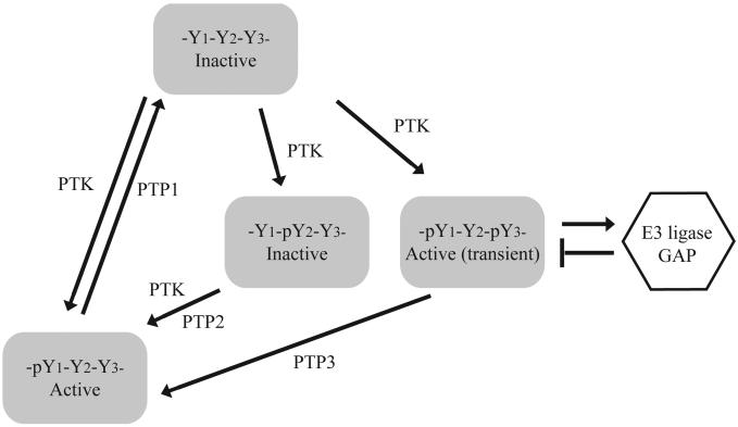
Positive and negative roles of tyrosine phosphorylation in cell signaling. In this illustration, three tyrosine residues (Y1, Y2, Y3) on a protein may be subject to phosphorylation by a PTK. Phosphorylation of Y1 increases the activity of the protein. Phosphorylation of Y2 inhibits the activity of the protein. Phosphorylation of Y3 induces feedback inhibition such as recruitment of E3 ligase that causes degradation of the protein or GTPase Activator Protein (GAP) that turns off G-proteins. While dephosphorylation of Y1 by PTP1 inactivate the protein, dephosphorylation of Y2 and Y3 by PTP2 and PTP3 are necessary for sustained activity of the protein. Thus, PTP1 is a negative regulator whereas PTP2 and PTP3 are positive regulators that coordinately control the activity of the protein.
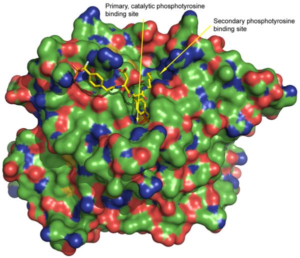
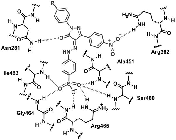
Illustration of the secondary phosphotyrosine binding site of PTP1B (from pdb code 1PXH), colored by element: carbon, green; oxygen, red; nitrogen blue, sulfur, yellow.
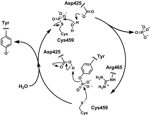
General mechanism of tyrosine dephosphorylation using Shp2 residue numbering. The nucleophylic cysteine residue Cys459, part of the signature PTP loop sequence (residues 457-467), VHCSAGIGRTG, attacks the phosphorus atom to break the PO bond in an SN2-like fashion. The aspartic acid residue Asp 425, that forms part of the WPD loop, acts as the proton donor to protonate the tyrosine phenol. This generates a labile thiophosphate intermediate that is subsequently hydrolysed to liberate the free cysteine residue. The aspartic acid plays a role as a general base, by abstracting a proton from the water molecule. The phosphotyrosine binding site of PTPs is generally populated with positively charged residues to stabilize the signature, in particular the arginine residue Arg465 of the P loop is important in stabilizing the negative charge of the pentavalent phosphorus formed upon initial addition of the cysteine to the phosphotyrosine group.
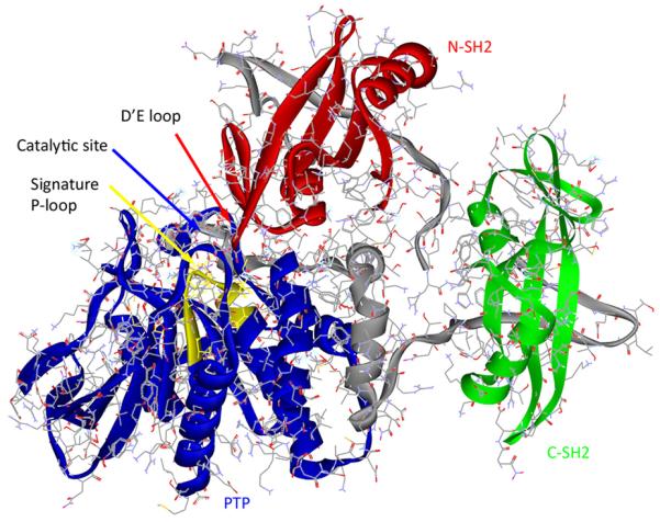
X-ray crystal structure of Shp2 (pdb code 2SHP) showing the N-SH2 (red), C-SH2 (green) and PTP (blue) domains. The two linking sequences that join these three domains are shown in gray. The signature P-loop motif (VHCSAGIGRTG) is illustrated in yellow.
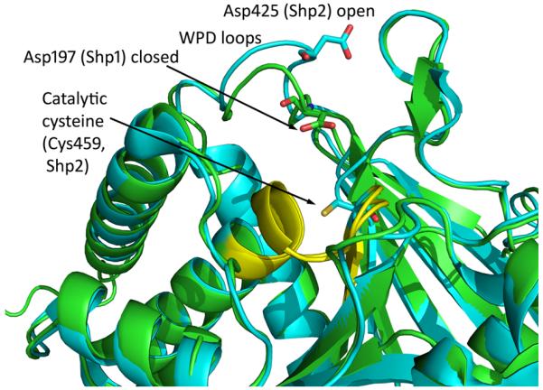
Overlay of the X-ray crystal structures of the PTP domain of Shp1 (pdb 1GWZ, colored green) and Shp2 (pdb 3B7O) colored blue, clearly indicating the closer proximity of the WPD aspartic acid residue to the catalytic cysteine residue for Shp1 as a consequence f the closed WPD loop conformation.
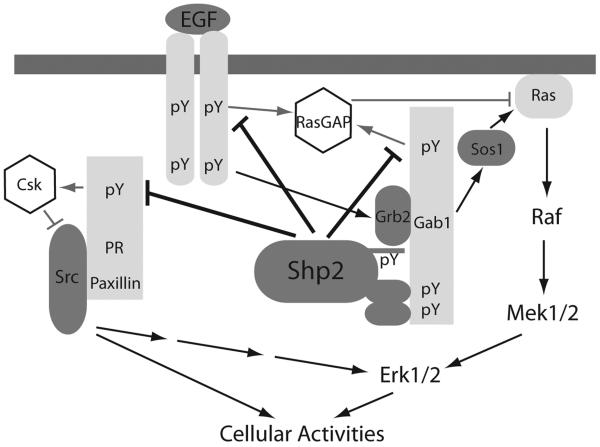
Signaling pathways from the EGF receptor to Erk1/2 and Src in adherent cells. Representative mechanisms are shown to illustrate that Shp2 activates Src family kinases by preventing recruitment of Csk to Src complexes and activates Ras-Erk1/2 by regulating RasGAP and Src.
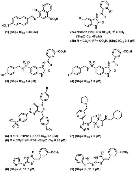
Representative of Shp2 inhibitors. (1), NSC-87877. (3) and (4) are synthesized based on screening hit (2). (5) and (6), PHPS1 and PHPS4.
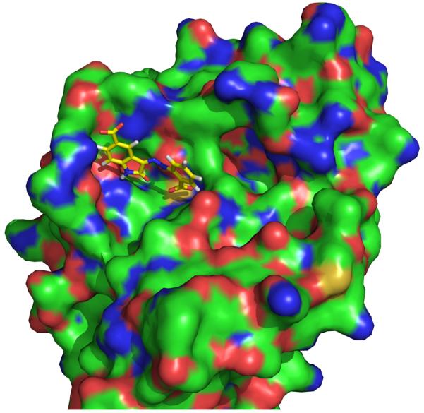
Docked structure of isatin (2b) with Shp2 (derived from structure pdb code 2SHP), colored by element: carbon, green; oxygen, red; nitrogen blue, sulfur, yellow: hydrogens removed from the protein for clarity.
Proposed schematic docking model of PHPS1 (5) with the catalytic site of Shp2. Hydrogen bonds between the ligand and phosphatase as predicted by MOE v2006.3 are shown as hatched lines.
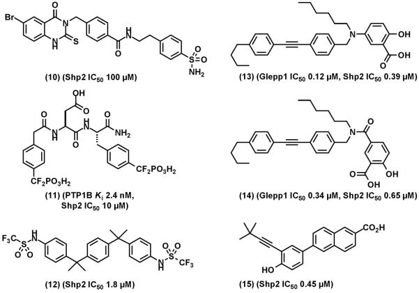
Shp2 inhibitors and other PTP inhibitors that cross-inhibit Shp2. (10), an Shp2 selective inhibitor identified from virtual screening of secondary binding site. (11), a potent PTP1B that cross inhibits Shp2. (13) and (14), orally bioavailable Glepp1 inhibitors that cross-inhibits Shp2. (15), a nuclear receptor small heterodimer partner that cross inhibits Shp2.
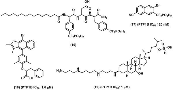
Representative of PTP1B inhibitors. (16), a cell permeable analog of compound (11). (17), an orally bioavailable PTP1B inhibitor. (18), Ertiprotafib. (19) MSI-1436 (Trodusquemine)
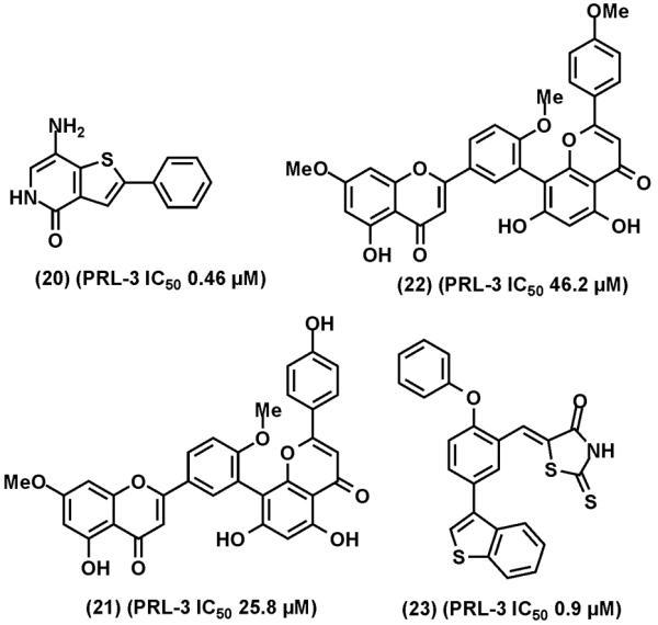
Representative of PRL-3 inhibitors. (20), thienopyridone. (21), ginkgetin. (22), sciadoptysin. (23), a rhodanine derivative.
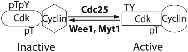
Cdc25 dual specificity phosphatases are positive regulators of Cdks. The activity of a Cdk is regulated by three phosphorylation sites Thr14, Tyr15, and Thr161 (residues numbers based on Cdk1). Thr14 and Tyr15 phosphorylation catalyzed by Wee1 and Myt1 kinases are inactivating while Thr161 phosphorylation by Cak is activating. Cdc25 phosphatases dephosphorylate Thr14 and Tyr15, resulting in Cdk activation.
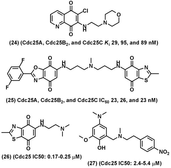
Representative of Cdc25 inhibitors. (24), NSC-663284. (25), IRC-083864, (26), BN82002, (27) BN82685.
References
-
- Hanahan D, Weinberg RA. The hallmarks of cancer. Cell. 2000;100:57–70. - PubMed
-
- Ostman A, Hellberg C, Bohmer FD. Protein-tyrosine phosphatases and cancer. Nat Rev Cancer. 2006;6:307–20. - PubMed
-
- Tonks NK. Protein tyrosine phosphatases: from genes, to function, to disease. Nat Rev Mol Cell Biol. 2006;7:833–46. - PubMed
Publication types
MeSH terms
Substances
Grants and funding
LinkOut - more resources
Full Text Sources
Other Literature Sources
