. 2010 Jun 1;59(1):71-80.
doi: 10.1111/j.1574-695X.2010.00663.x. Epub 2010 Feb 17.

gammadelta T cells promote the maturation of dendritic cells during West Nile virus infection

Affiliations

gammadelta T cells promote the maturation of dendritic cells during West Nile virus infection

Hao Fang et al. FEMS Immunol Med Microbiol. .

Abstract

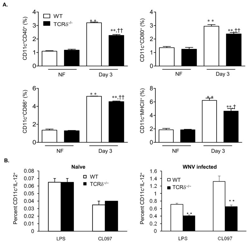
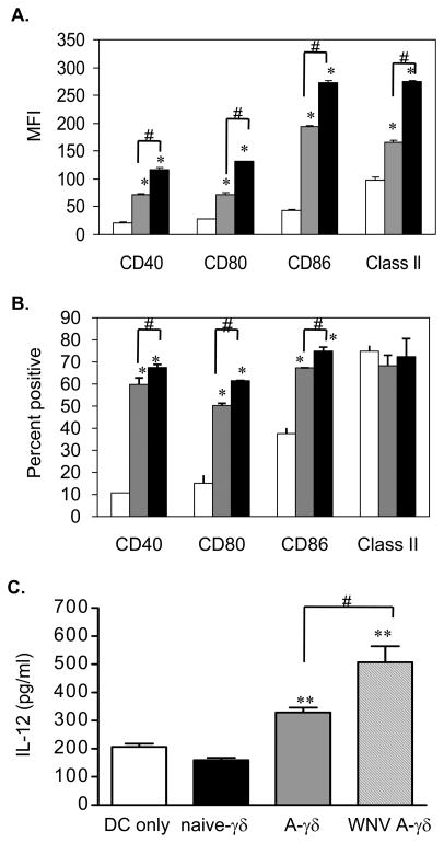
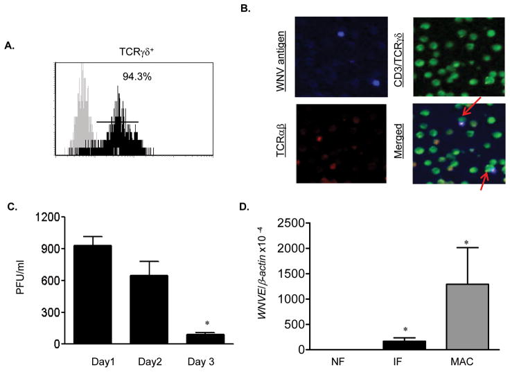
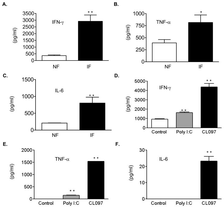
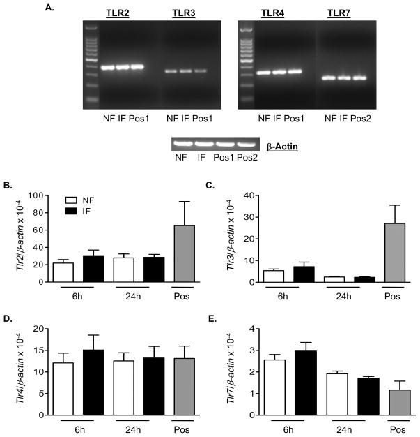
gammadelta T cells are important for the early control of West Nile virus (WNV) dissemination. Here, we investigated the role of gammadelta T cells in the regulation of CD4(+) T-cell response following a WNV challenge. Splenic dendritic cells (DCs) of WNV-infected gammadelta T-cell-deficient (TCRdelta(-/-)) mice displayed lower levels of CD40, CD80, CD86 and major histocompatibility complex (MHC) class II expression and interleukin-12 (IL-12) production than those of wild-type mice. Naïve DCs cocultured with WNV-infected gammadelta T cells showed enhanced levels of costimulatory molecules, MHC class II expression and IL-12 production. Further, coculture of CD4(+) T cells from OT II transgenic mice with DCs of WNV-infected TCRdelta(-/-) mice induced less interferon-gamma (IFN-gamma) and IL-2 production than with those of wild-type controls. Viral antigens were detected in WNV-infected gammadelta T cells.WNV infection or toll-like receptor (TLR) agonist treatment of gammadelta T cells induced the production of IFN-gamma, tumor necrosis factor-alpha and IL-6, which are known to promote DC maturation. Nevertheless, the levels of TLRs 2, 3, 4 and 7 expression of WNV-infected gammadelta T cells were not different from those of noninfected cells. Overall, these data suggest that WNV-induced gammadelta T-cell activation promotes DC maturation and initiates CD4(+) T-cell priming.


Figures

Figure 1
Figure 1
DC maturation in WNV-infected mice. A, The percentages of CD40+ CD11c+, CD80+CD11c+, CD86+CD11c+ or MHCII+CD11c+ in the spleens. Data are presented as means ± SEM, n = 4 or 5. ** P < 0.01 for WNV-infected (IF) vs. non-infected (NF). P < 0.05 or †† P < 0.01 for wild-type (WT) vs. TCRδ−/− mice. B, The percentages of IL-12+ CD11c+ naïve (left panel) or day 2 WNV-infected (right panel) splenocytes after ex vivo stimulation with LPS or CL097. Data are presented as means ± SEM, n = 4 or 5. ** P < 0.01 for wild-type vs. TCRδ−/− mice. P values were calculated with a non-paired Student’st test.
Figure 2
Figure 2
In vitro DC maturation assay. Cell surface molecule expression of CD11c+ cells alone (white bar) or co-culture with γδ T cells of naïve (grey bar) or WNV-infected (black bar) mice as determined by MFI (A) and the percentage (B). C, IL-12 production from co-culture of naïve DCs with in vitro WNV infected γδ T cells measured by ELISA. A-γδ: anti-CD3 activated γδ T cells. WNV A-γδ: anti-CD3 treated γδ T cells infected with WNV. Data are presented as means ± SEM, n = 6. *P < 0.05 or ** P < 0.01 for co-cultured vs. DC alone. #P < 0.05 for IF vs. NF. P values were calculated with a non-paired Student’st test. Results are representative of two independent experiments.
Figure 3
Figure 3
In vitro WNV infection of γδ T cells. A, Splenic γδ T cells were stained with antibody to γδ and analyzed by flow cytometer. Dark area represents cells stained with anti-γδ; gray area, unstained cells. B, Immunofluorescence photomicrographs of WNV-infected γδ T cells at day 2 post-infection. Infected cells were stained for WNV antigen (blue), CD3/TCRγδ (green) and TCRαβ (red). Arrows point to CD3+/γδ+WNV+ cells. C, Plaque assay of virus titer. *P < 0.05 for day 3 vs. day 1. D, WNV infection in splenic γδ T cells as measured by Q-PCR. Negative and positive controls represent uninfected γδ T cells and WNV-infected H36.12j cells (MAC), respectively. *P < 0.05 for non-infected vs. WNV-infected cells. P values were calculated with a non-paired Student’st test.
Figure 4
Figure 4
WNV infection or TLR agonist stimulation of γδ T cells induces proinflammatory cytokines. A–C, Supernatant was collected at day 4 post-infection. D–F, Supernatant was collected 24 h post-treatment. Data are presented as means ± SEM, n = 6. * P < 0.05 or ** P < 0.01 for non-infected or non-treated vs. WNV-infected or treated. P values were calculated with a non-paired Student’st test.
Figure 5
Figure 5
Levels of TLR expression on WNV-infectedγδ T cells. Splenic γδ T cells were infected with WNV and harvested at indicated time points. A, PCR amplification of TLRs (top panel) or β-actin (bottom panel) of WNV-infected or non-infected γδ T cells harvested at 24 h post-infection. B–E, Q-PCR analysis of TLRs. B, TLR2. C, TLR3. D, TLR4. E, TLR7. cDNA of CD11c+ cells or plasmacytoid DCs were used as positive control 1 & 2. Data are presented as means ± SEM, n = 7 or 8. P values were calculated with a Mann-Whitney test.

Similar articles

Cited by

References

    1. Anderson JF, Rahal JJ. Efficacy of interferon alpha-2b and ribavirin against West Nile virus in vitro. Emerg Infect Dis. 2002;8:107–108. - PMC - PubMed
    1. Argentati K, Re F, Donnini A, et al. Numerical and functional alterations of circulating gammadelta T lymphocytes in aged people and centenarians. J Leukoc Biol. 2002;72:65–71. - PubMed
    1. Beetz S, Marischen L, Kabelitz D, Wesch D. Human gamma delta T cells: candidates for the development of immunotherapeutic strategies. Immunol Res. 2007;37:97–111. - PubMed
    1. Beetz S, Wesch D, Marischen L, Welte S, Oberg HH, Kabelitz D. Innate immune functions of human gammadelta T cells. Immunobiology. 2008;213:173–182. - PubMed
    1. Bennett SR, Carbone FR, Karamalis F, Flavell RA, Miller JF, Heath WR. Help for cytotoxic-T-cell responses is mediated by CD40 signalling. Nature. 1998;393:478–480. - PubMed

Publication types