. 2010 Mar 25:11:14.
doi: 10.1186/1471-2091-11-14.

Interaction of connexin43 and protein kinase C-delta during FGF2 signaling

Affiliations

Interaction of connexin43 and protein kinase C-delta during FGF2 signaling

Corinne Niger et al. BMC Biochem. .

Abstract

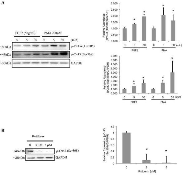
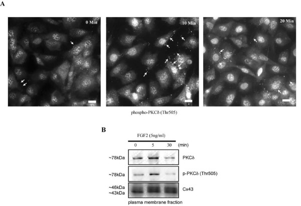
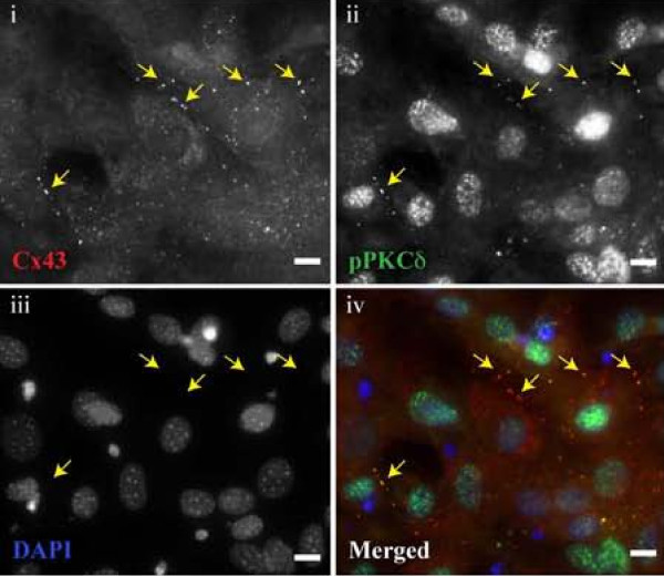
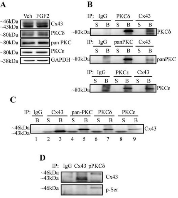
Background: We have recently demonstrated that modulation of the gap junction protein, connexin43, can affect the response of osteoblasts to fibroblast growth factor 2 in a protein kinase C-delta-dependent manner. Others have shown that the C-terminal tail of connexin43 serves as a docking platform for signaling complexes. It is unknown whether protein kinase C-delta can physically interact with connexin43.

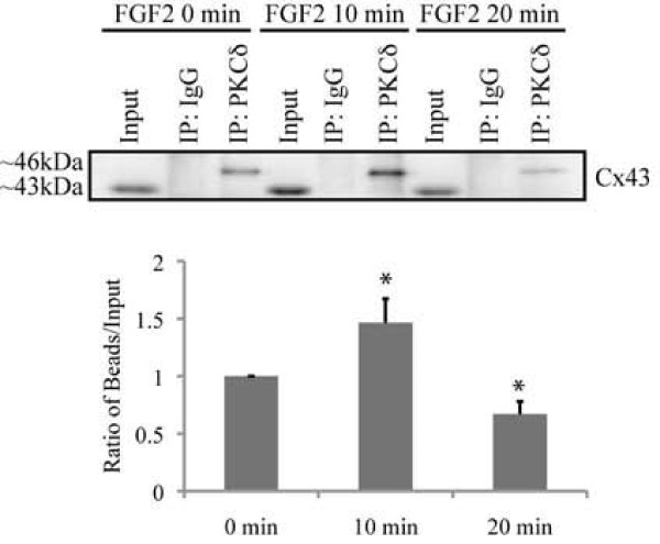
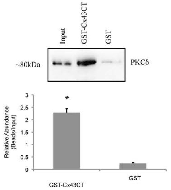
Results: In the present study, we investigate by immunofluorescent co-detection and biochemical examination the interaction between Cx43 and protein kinase C-delta. We establish that protein kinase C-delta physically interacts with connexin43 during fibroblast growth factor 2 signaling, and that protein kinase C delta preferentially co-precipitates phosphorylated connexin43. Further, we show by pull down assay that protein kinase C-delta associates with the C-terminal tail of connexin43.

Conclusions: Connexin43 can serve as a direct docking platform for the recruitment of protein kinase C-delta in order to affect fibroblast growth factor 2 signaling in osteoblasts. These data expand the list of signal molecules that assemble on the connexin43 C-terminal tail and provide a critical context to understand how gap junctions modify signal transduction cascades in order to impact cell function.


Figures

Figure 1
Figure 1
Phosphorylation of Cx43 at serine 368 by PKCδ. (A). Western blots (n = 5) performed with anti-phospho-PKCδ (Thr505) and anti-phospho-Cx43 (Ser368) antibodies show concomitant phosphorylation of PKCδ and Cx43 following treatment with 5 ng/ml FGF2 and 200 nM PMA (positive control). (B). Western blots (n = 3) probed with anti-phospho-Cx43 (Ser368) antibodies reveal that the PKCδ inhibitor, rottlerin (3 μM and 5 μM), can block the FGF2-induced phosphorylation of Cx43 at serine 368. Blots probed with anti-GAPDH antibodies are to verify equal loading. Representative blots are shown. Mean densitometric values of the blots from all experiments are shown in the accompanying bar graphs. Error bars indicate standard deviation. An asterisk denotes a p-value <0.05, relative to control.
Figure 2
Figure 2
FGF2 induces the accumulation of phospho-PKCδ in both the nuclei and plasma membrane. (A). Serum starved MC3T3 cells were subjected to immunofluorescent detection with anti-phospho-PKCδ (Thr505) antibodies during a time course of treatment with FGF2 (5 ng/ml). Representative images collected by epi-fluorescence microscopy are shown for each time point. Small arrows indicate the punctate staining at the cell periphery. The scale bar indicates ~10 μm. (B) Western blots (n = 3) probed with anti-PKCδ, anti-phospho-PKCδ (Thr505) and anti-Cx43 antibodies were performed on equal amounts of plasma membrane protein extracts from FGF2 (5 ng/ml) treated osteoblasts. Representative blots are shown.
Figure 3
Figure 3
Co-detection of Cx43 and phospho-PKCδ at the plasma membrane. Serum starved MC3T3 cells were treated with FGF2 for 10 min prior to fixation and subsequent staining with (i) mouse anti-Cx43 (red), (ii) rabbit anti-phospho-PKCδ (Thr505) (green), (iii) DAPI (blue). A merged image is also shown (iv). Arrows indicate the same position on each panel and correspond to areas where both Cx43 and phospho-PKCδ appear to be closely co-detected in their respective panels. A representative field of view is shown. The white scale bar is ~10 μm.
Figure 4
Figure 4
Connexin43 and PKCδ are present in a protein complex. (A). Whole cell extracts from cells treated with vehicle or 5 ng/ml FGF2 for 15 minutes were subjected to western blotting with the indicated antibodies (n = 3). (B). Whole cell extracts were immunoprecipitated (IP) with non-immune IgG, anti-PKC (δ, pan or ε) or anti-Cx43 antibodies, as indicated. The supernatant (S) and bead (B) fractions from the immunoprecipitations were subjected to western blotting (WB) with anti-PKC (δ, pan or ε) antibodies. Immunoprecipitation with anti-Cx43 antibodies co-precipitates PKCδ (as well as PKCε). (C). Whole cell extracts were immunoprecipitated with non-immune IgG, anti-Cx43, or anti-PKC (pan, δ, or ε) antibodies, as indicated. The supernatant (S) and bead (B) fractions from the immunoprecipitations were subjected to western blotting with Cx43 antibodies. All three anti-PKC antibodies co-precipitate Cx43. (D). Whole cell extracts were prepared from cells treated with 5 ng/ml FGF2 for 10 minutes. The extracts were immunoprecipitated with non-immune IgG, anti-Cx43, or anti-phospho-PKCδ (Thr505) antibodies, as indicated. The bead fractions from the immunoprecipitations were subjected to western blotting with anti-Cx43 or anti-phospho-serine antibodies. Anti-phospho-PKCδ (Thr505) antibodies preferentially co-immunoprecipitate serine phosphorylated Cx43.
Figure 5
Figure 5
FGF2 treatment promotes a transient increase in association of Cx43 and phospho-PKCδ. Whole cell extracts were prepared from cells treated with vehicle or 5 ng/ml FGF2 for the indicated time. The extracts were immunoprecipitated (IP) with non-immune IgG, anti-Cx43, or anti-phospho-PKCδ (Thr505) antibodies, as indicated. A fraction of the input and the bead fractions from the immunoprecipitations were subjected to western blotting (WB) with anti-Cx43 antibodies. The co-precipitated Cx43 and phospho-PKCδ complex is most abundant at 10 minutes and returns to basal levels by 15 minutes. The graph reveals the average ratio of co-immunoprecipitated Cx43 relative to the Cx43 present in the input fraction for each time point from three separate experiments. Error bars indicate standard deviation. An asterisk denotes a p-value <0.05, relative to control. The ratio for the 0 min FGF2 treatment point has been arbitrarily set to a value of 1.
Figure 6
Figure 6
PKCδ associates with the C-terminal tail of Cx43. GST pull downs were performed using GST- or GST-Cx43CT(241-381) crosslinked beads and whole cell extracts from MC3T3 cells. The input, and bead fractions were analyzed by western blot. The blots were probed with anti-PKCδ antibodies. PKCδ is pulled down by the GST-Cx43CT(241-381) construct but not by GST-crosslinked beads alone. Mean densitometric values of the blots are shown in the accompanying bar graph. The graph reveals the ratio of "pulled-down" PKCδ relative to the PKCδ present in the input fraction from three separate experiments. Error bars indicate standard deviation. An asterisk denotes a p-value <0.05, relative to control.

Similar articles

Cited by

References

    1. Stains JP, Civitelli R. Gap junctions in skeletal development and function. Biochim Biophys Acta. 2005;1719(1-2):69–81. doi: 10.1016/j.bbamem.2005.10.012. - DOI - PubMed
    1. Jiang JX, Siller-Jackson AJ, Burra S. Roles of gap junctions and hemichannels in bone cell functions and in signal transmission of mechanical stress. Front Biosci. 2007;12:1450–1462. doi: 10.2741/2159. - DOI - PMC - PubMed
    1. Laird DW. Life cycle of connexins in health and disease. Biochem J. 2006;394(Pt 3):527–543. - PMC - PubMed
    1. Wei CJ, Xu X, Lo CW. Connexins and cell signaling in development and disease. Annu Rev Cell Dev Biol. 2004;20:811–838. doi: 10.1146/annurev.cellbio.19.111301.144309. - DOI - PubMed
    1. Gerido DA, White TW. Connexin disorders of the ear, skin, and lens. Biochim Biophys Acta. 2004;1662(1-2):159–170. doi: 10.1016/j.bbamem.2003.10.017. - DOI - PubMed

Publication types