. 2010 Jul 15;49(2):147-58.
doi: 10.1016/j.freeradbiomed.2010.03.017. Epub 2010 Mar 23.

Triterpenoids CDDO-ethyl amide and CDDO-trifluoroethyl amide improve the behavioral phenotype and brain pathology in a transgenic mouse model of Huntington's disease

Affiliations

Triterpenoids CDDO-ethyl amide and CDDO-trifluoroethyl amide improve the behavioral phenotype and brain pathology in a transgenic mouse model of Huntington's disease

Cliona Stack et al. Free Radic Biol Med. .

Abstract

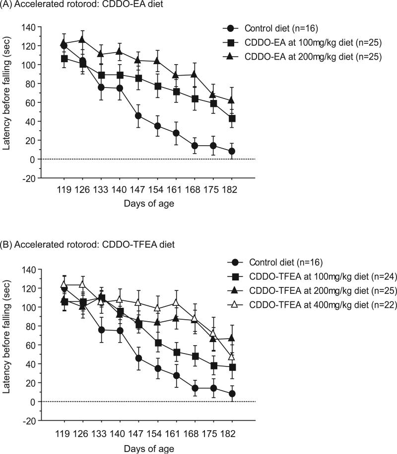
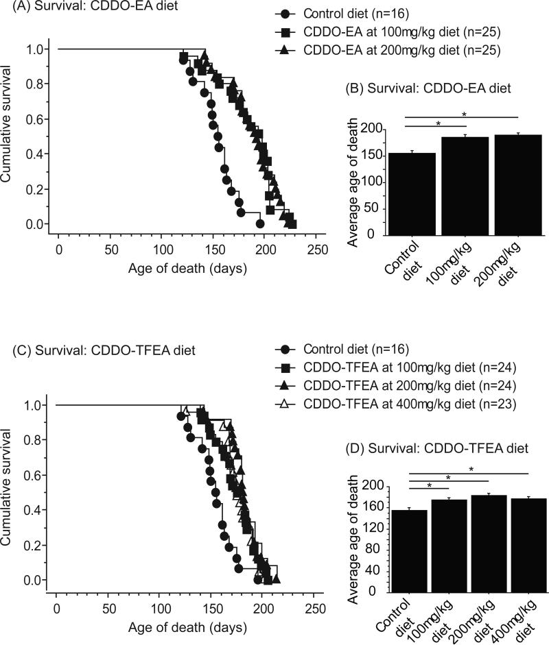
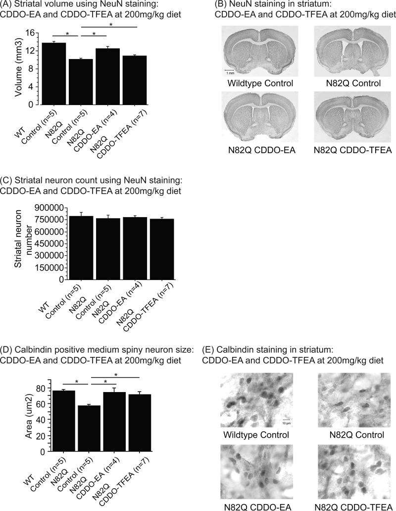
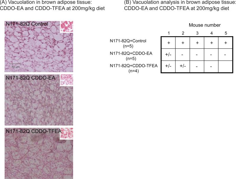
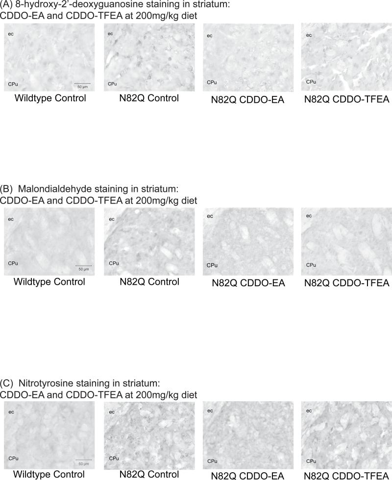
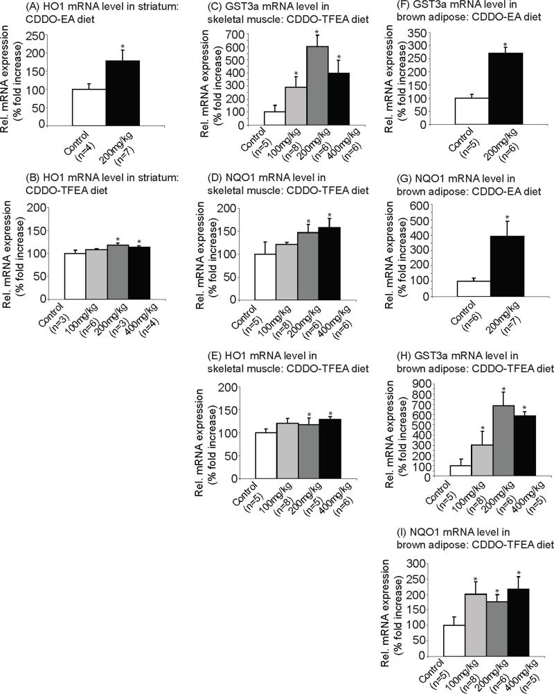
Oxidative stress is a prominent feature of Huntington's disease (HD) due to mitochondrial dysfunction and the ensuing overproduction of reactive oxygen species (ROS). This phenomenon ultimately contributes to cognitive and motor impairment, as well as brain pathology, especially in the striatum. Targeting the transcription of the endogenous antioxidant machinery could be a promising therapeutic approach. The NF-E2-related factor-2 (Nrf2)/antioxidant response element (ARE) signaling pathway is an important pathway involved in antioxidant and anti-inflammatory responses. Synthetic triterpenoids, which are derived from 2-Cyano-3,12-Dioxooleana-1,9-Dien-28-Oic acid (CDDO) activate the Nrf2/ARE pathway and reduce oxidative stress in animal models of neurodegenerative diseases. We investigated the effects of CDDO-ethyl amide (CDDO-EA) and CDDO-trifluoroethyl amide (CDDO-TFEA) in N171-82Q mice, a transgenic mouse model of HD. CDDO-EA or CDDO-TFEA were administered in the diet at various concentrations, starting at 30days of age. CDDO-EA and CDDO-TFEA upregulated Nrf2/ARE induced genes in the brain and peripheral tissues, reduced oxidative stress, improved motor impairment and increased longevity. They also rescued striatal atrophy in the brain and vacuolation in the brown adipose tissue. Therefore compounds targeting the Nrf2/ARE pathway show great promise for the treatment of HD.


Figures

Figure 1
Figure 1. CDDO-ethyl amide (CDDO-EA) and CDDO-trifluoroethyl amide (CDDO-TFEA) improved motor performance in N171-82Q mice
(A) Latency to fall during rotorod trials of N171-82Q mice treated with control diet, CDDO-EA at 100mg/kg diet and 200mg/kg diet. Data were expressed as means and standard errors. N171-82Q mice treated with CDDO-EA 100mg/kg diet and CDDO-EA 200mg/kg diet both showed significantly improved motor coordination compared to N171-82Q mice fed with control diet (p=0.04, p=0.0006). (B) Latency to fall during rotorod trials of N171-82Q mice treated with control diet, CDDO-TFEA at 100mg/kg diet, 200mg/kg diet and 400mg/kg diet. Data were expressed as means and standard errors. N171-82Q mice treated with CDDO-TFEA at 200mg/kg diet and CDDO-TFEA 400mg/kg diet both showed significantly improved motor coordination compared to N171-82Q mice fed control diet (p=0.005, p=0.0007).
Figure 2
Figure 2. CDDO-ethyl amide (CDDO-EA) and CDDO-trifluoroethyl amide (CDDO-TFEA) improved survival in N171-82Q mice
(A) Cumulative survival and (B) average age of death of N171-82Q mice treated with control diet, CDDO-EA at 100mg/kg diet and 200mg/kg diet. Data were expressed as means and standard errors. N171-82Q mice treated with CDDO-EA 100mg/kg diet and CDDO-EA 200mg/kg diet both showed significantly improved survival compared to mice fed control diet (p=0.0002, p<0.0001). (C) Cumulative survival and (D) average age of death of N171-82Q mice treated with control diet, CDDO-TFEA at 100mg/kg diet, 200mg/kg diet and 400mg/kg diet. Data were expressed as means and standard errors. N171-82Q mice treated with CDDO-TFEA at 100mg/kg diet, CDDO-TFEA 200mg/kg diet and CDDO-TFEA 400mg/kg diet each showed significantly improved survival compared to control (p=0.001, p=<0.0001, p=0.0003).
Figure 3
Figure 3. CDDO-ethyl amide (CDDO-EA) and CDDO-trifluoroethyl amide (CDDO-TFEA) attenuated striatal atrophy and reduction of medium spiny neuron size in N171-82Q mice
(A) Striatal volume of NeuN labeled tissues of N171-82Q mice treated with control diet, CDDO-EA and CDDO-TFEA at 200mg/kg diet. Data were expressed as means and standard errors. (B) Photographs of brain sections labeled with NeuN antibody. N171-82Q mice fed control diet showed a significant reduction in striatal volume compared to wildtype mice fed control diet (p<0.0001). However, treatment with both CDDO-EA and CDDO-TFEA significantly increased striatal volume in N171-82Q mice compared to N171-82Q mice fed control diet (p<0.0001, p=0.002). (C) Striatal neuron count in NeuN labeled tissues of N171-82Q mice treated with control diet, CDDO-EA and CDDO-TFEA at 200mg/kg diet. Data were expressed as means and standard errors. There was no difference in striatal neuron count between any of the groups. (D) Calbindin positive medium spiny neuron size of N171-82Q mice treated with control diet, CDDO-EA and CDDO-TFEA at 200mg/kg diet. Data were expressed as means and standard errors. (E) Photographs of brain sections labeled with calbindin antibody. N171-82Q mice fed control diet showed a significant reduction in medium spiny neuron size compared to wildtype mice fed control diet (p=0.001). N171-82Q mice treated with CDDO-EA and CDDO-TFEA showed significantly larger medium spiny neuron size compared to N171-82Q mice fed control diet (p=0.004, p=0.006).
Figure 4
Figure 4. CDDO-ethyl amide (CDDO-EA) and CDDO-trifluoroethyl amide (CDDO-TFEA) reduced vacuolation in the brown adipose tissue of N171-82Q mice
(A) Photographs of brown adipose tissue stained by hematoxylin and eosin in N171-82Q transgenic mice fed control diet, CDDO-EA and CDDO-TFEA at 200mg/kg diet. Right insets show the red oil staining for lipids. (B) Vacuolation analysis of the brown adipose tissue in N171-82Q transgenic mice fed control diet, CDDO-EA and CDDO-TFEA at 200mg/kg diet. Data were expressed as “-” for the lack of extensive vacuolation, “+” for the presence of extensive vacuolation, and “+/-” for the presence of vacuolation in few area.
Figure 5
Figure 5. CDDO-ethyl amide (CDDO-EA) and CDDO-trifluoroethyl amide (CDDO-TFEA) reduces oxidative damage in N171-82Q mice
(A) 8-hydroxy-2’-deoxyguanosine staining in striatal tissue of mice treated with control diet, CDDO-EA and CDDO-TFEA at 200mg/kg diet. (B) Malondialdehyde staining in striatal tissue of mice treated with control diet, CDDO-EA and CDDO-TFEA at 200mg/kg diet. (C) 3-Nitrotyrosine staining in striatal tissue of mice treated with control diet, CDDO-EA and CDDO-TFEA at 200mg/kg diet. Oxidative stress was increased in N171-82Q mice fed control diet compared to wildtype mice fed control diet. Administration of CDDO-EA and CDDO-TFEA in N171-82Q mice reduced oxidative stress compared to N171-82Q mice fed control diet.
Figure 6
Figure 6. CDDO-ethyl amide (CDDO-EA) and CDDO-trifluoroethyl amide (CDDO-TFEA) upregulated expression of genes specifically involved in the Nrf2/ARE pathway in N171-82Q mice
mRNA levels of HO1 in striatum of (A) CDDO-EA treated and (B) CDDO-TFEA treated N171-82Q mice. Data were expressed as means and standard errors. HO1 gene expression was significantly increased in striatum of N171-82Q mice treated with CDDO-EA (p=0.04 at 200mg/kg diet) and CDDO-TFEA (p=0.009 at 200mg/kg diet, p=0.03 at 400mg/kg diet) compared to N171-82Q mice fed control diet. mRNA levels of (C) GST3a (D) NQO1 and (E) HO1 in skeletal muscle of CDDO-TFEA treated N171-82Q mice. Data were expressed as means and standard errors. mRNA level of GST3a (p=0.007 at 100mg/kg diet, p=0.0001 at 200mg/kg diet, p=0.002 at 400mg/kg diet), NQO1 (p=0.04 at 200mg/kg diet, p=0.02 at 400mg/kg diet) and HO1 (p=0.02 at 200mg/kg diet, p=0.03 at 400mg/kg diet) were significantly increased compared to mice fed control diet. mRNA levels of (F) GST3a and (G) NQO1 in brown adipose tissue of CDDO-EA treated N171-82Q mice. Data were expressed as means and standard errors. GST3a gene expression was significantly increased in brown adipose tissue of CDDO-EA treated mice compared to control (p=0.005 at 200mg/kg diet). NQO1 gene expression was also significantly increased in brown adipose tissue of CDDO-EA treated mice compared to control (p=0.0002 at 200mg/kg diet). mRNA levels of (H) GST3a and (I) NQO1 in brown adipose tissue of CDDO-TFEA treated N171-82Q mice. Data were expressed as means and standard errors. Both GST3a gene expression (p=0.003 at 100mg/kg diet, p<0.0001 at 200mg/kg diet, p<0.0001 at 400mg/kg diet) and NQO1 gene expression (p=0.005 at 100mg/kg diet, p=0.03 at 200mg/kg diet, p=0.005 at 400mg/kg diet) were significantly increased in CDDO-TFEA treated mice.

Comment in

References

    1. Halliday GM, McRitchie DA, Macdonald V, Double KL, Trent RJ, McCusker E. Regional specificity of brain atrophy in Huntington's disease. Exp Neurol. 1998;154(2):663–672. - PubMed
    1. Vonsattel JP, Myers RH, Stevens TJ, Ferrante RJ, Bird ED, Richardson EP., Jr Neuropathological classification of Huntington's disease. J Neuropathol Exp Neurol. 1985;44(6):559–577. - PubMed
    1. Ferrante RJ, Kowall NW, Richardson EP., Jr Proliferative and degenerative changes in striatal spiny neurons in Huntington's disease: a combined study using the section-Golgi method and calbindin D28k immunocytochemistry. J Neurosci. 1991;11(12):3877–3887. - PMC - PubMed
    1. Sorolla MA, Reverter-Branchat G, Tamarit J, Ferrer I, Ros J, Cabiscol E. Proteomic and oxidative stress analysis in human brain samples of Huntington disease. Free Radic Biol Med. 2008;45(5):667–678. - PubMed
    1. Browne SE, Beal MF. Oxidative damage in Huntington's disease pathogenesis. Antioxid Redox Signal. 2006;8(11-12):2061–2073. - PubMed

Publication types

MeSH terms

Substances