. 2010 Jul 1;51(3):1242-52.
doi: 10.1016/j.neuroimage.2010.03.040. Epub 2010 Mar 23.

High-resolution structural and functional MRI of hippocampal CA3 and dentate gyrus in patients with amnestic Mild Cognitive Impairment

Affiliations

High-resolution structural and functional MRI of hippocampal CA3 and dentate gyrus in patients with amnestic Mild Cognitive Impairment

Michael A Yassa et al. Neuroimage. .

Abstract

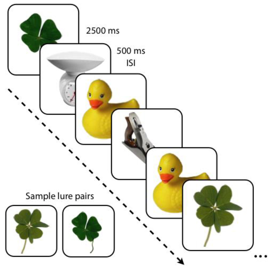
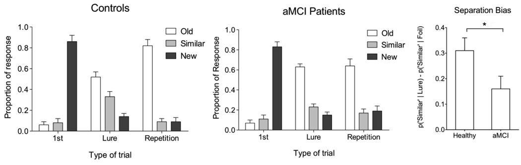
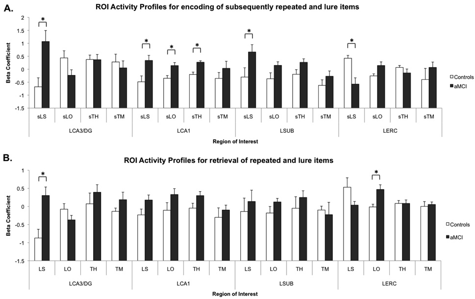
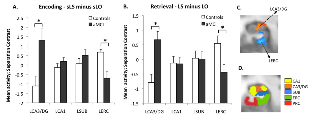
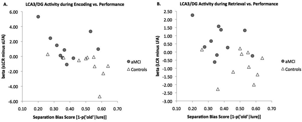
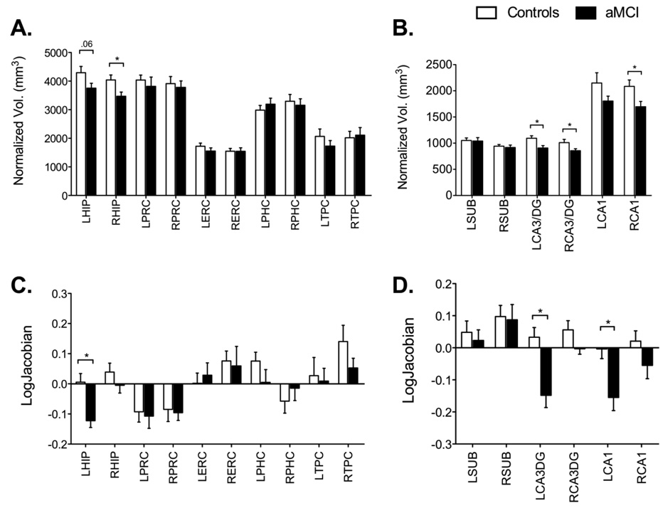
Functional magnetic resonance imaging (fMRI) studies have observed hyperactivity in the hippocampal region in individuals with Mild Cognitive Impairment (MCI). However, the actual source of such hyperactivity is not well understood. Studies of aged rats observed similar hyperactive signals in the CA3 region of the hippocampus that correlated with spatial memory deficits and, in particular, with their ability to represent novel environments as being distinct from familiar ones (pattern separation). In this study, we tested the hypothesis that patients with amnestic MCI (aMCI) have deficits in pattern separation, along with hyperactive fMRI BOLD activity in the CA3 region of the hippocampus. We used high-resolution fMRI during a continuous recognition task designed to emphasize pattern separation. We conducted hippocampal subfield-level region of interest analyses to test for dysfunctional activity in aMCI patients. We found that patients showed impaired performance on trials that taxed their pattern separation abilities. We also observed hyperactive BOLD signals in the CA3/dentate and hypoactive signals in the entorhinal cortex during the separation condition. In a high-resolution morphometric analysis of hippocampal subfields, aMCI patients also had smaller CA3/dentate and CA1 volumes (no difference in the subiculum). The CA3/dentate region bilaterally also exhibited the largest shape deformations in aMCI patients, suggesting that this locus is affected early in the course of the disease. These findings suggest that structural and functional changes in the CA3/dentate region of the hippocampus contribute to the deficits in episodic memory that are observed in patients with aMCI. The functional hyperactivity may be evidence for a dysfunctional encoding mechanism, consistent with the predictions of computational models of hippocampal learning.


Figures

Figure 1
Figure 1
Behavioral task. Pictures of single items were presented for 2000 ms followed by a 500 ms ISI. Novel, repeated, and similar lure items were randomly shuffled in the task. A sample pair of similar stimuli used as lures is shown.
Figure 2
Figure 2
Behavioral data in the two groups. There are no significant differences across groups on the repeated items or new items (P>0.05). There is a significant difference across groups on the critical lure items, where aMCI patients are more likely to call these items “old” (63% vs. 52% for controls; P<0.05). The separation bias metric is another way to clearly show this difference. It is calculated as p(“Similar”|Lure)-p(“Similar”|Foil). There is a significant difference on this bias metric between groups (P<0.05). All bars are mean ± s.e.m.
Figure 3
Figure 3
Overall main effect across conditions showing generally increased activity throughout hippocampal subregions during encoding and retrieval. Panel A shows the four ROIs activity profiles during encoding of subsequently repeated and lure items. Of critical interest are the higher activity in MCI patients in multiple hippocampal subregions and the lower activity in the entorhinal cortex. Panel B shows the same ROIs activity profiles during retrieval of repeated and lure items. Here aMCI patients show significantly higher activity in the left CA3/DG during lure correct rejections and higher activity in the left entorhinal cortex during lure false alarms. In general, both these panels illustrate that there is a tendency towards higher activity in aMCI patients across conditions and ROIs, although most are not significant. All bars are mean ± s.e.m. Condition abbreviations: sLS = lures subsequently called “similar”, sLO = lures subsequently called “old”, sTH = target subsequent hits, sTM = target subsequent misses, LS = lures called “similar”, LO = lures called “old”, TH = target hits, TM = target misses.
Figure 4
Figure 4
Results of the critical contrasts. (A) During encoding (lures subsequently called “similar” minus lures subsequently called “old”) aMCI patients had significantly higher activity in the left CA3/DG and lower activity in the left entorhinal cortex. (B) During retrieval (lures called “similar” minus lures called “old”), there was an identical pattern of group difference. All bars are mean ± s.e.m. (C) The two ROIs of interest in this contrast during encoding shown on an average customized structural template based on the entire sample (functional ROIs based on the t-contrast between groups). Group difference in activity during retrieval was very similar. (D) A sample slice from the anatomical segmentation template in the left medial temporal lobe.
Figure 5
Figure 5
Correlations between hyperactivity in the left CA3/DG and behavioral performance during encoding (A) and during retrieval (B). There is a significant negative correlation between performance and hyperactivity during both encoding and retrieval.
Figure 6
Figure 6
Volumetric and morphometric analyses. Panel A shows that only the hippocampus underwent volumetric changes in the aMCI patients. Panel B shows a more detailed analysis of hippocampal subfields and shows that volumetric decreases were limited to the CA3/DG and CA1 subfields only and not the subiculum. Panel C shows again that the left hippocampus is the only region that underwent morphometric (i.e., shape) changes in the aMCI patients. Panel D shows a detailed morphometric analysis of hippocampal subfields and shows that once again this change can be ascribed to the CA3/DG and CA1 subfields and not the subiculum. All bars are mean ± s.e.m.

Similar articles

Cited by

References

    1. Ances BM, Liang CL, Leontiev O, Perthen JE, Fleisher AS, Lansing AE, Buxton RB. Effects of aging on cerebral blood flow, oxygen metabolism, and blood oxygenation level dependent responses to visual stimulation. Human Brain Mapping. 2009;30(4):1120–1132. - PMC - PubMed
    1. Anderson ND, Ebert PL, Jennings JM, Grady CL, Cabeza R, Graham SJ. Recollection-and familiarity-based memory in healthy aging and amnestic mild cognitive impairment. Neuropsychology. 2008;22(2):177–187. - PubMed
    1. Baker CI, Hutchison TL, Kanwisher N. Does the fusiform face area contain subregions highly selective for nonfaces? Nature Neuroscience. 2007;10(1):3–4. - PubMed
    1. Bakker A, Kirwan CB, Miller NI, Stark CEL. Pattern separation in the human hippocampal CA3 and dentate gyrus. Science. 2008;319:1640–1642. - PMC - PubMed
    1. Bassett SS, Yousem DM, Cristinzio C, Kusevic I, Yassa MA, Caffo BS, Zeger SL. Familial risk for Alzheimer's disease alters fMRI activation patterns. Brain : A Journal of Neurology. 2006;129(Pt 5):1229–1239. - PMC - PubMed

Publication types