. 2010 May 28;285(22):17098-111.
doi: 10.1074/jbc.M110.109439. Epub 2010 Mar 25.

eIF2alpha phosphorylation tips the balance to apoptosis during osmotic stress

Affiliations

eIF2alpha phosphorylation tips the balance to apoptosis during osmotic stress

Elena Bevilacqua et al. J Biol Chem. .

Abstract

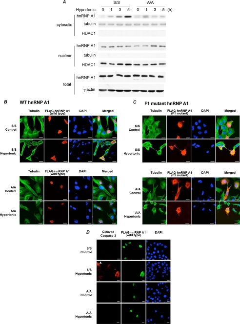
Regulation of cell volume is of great importance because persistent swelling or shrinkage leads to cell death. Tissues experience hypertonicity in both physiological (kidney medullar cells) and pathological states (hypernatremia). Hypertonicity induces an adaptive gene expression program that leads to cell volume recovery or apoptosis under persistent stress. We show that the commitment to apoptosis is controlled by phosphorylation of the translation initiation factor eIF2alpha, the master regulator of the stress response. Studies with cultured mouse fibroblasts and cortical neurons show that mutants deficient in eIF2alpha phosphorylation are protected from hypertonicity-induced apoptosis. A novel link is revealed between eIF2alpha phosphorylation and the subcellular distribution of the RNA-binding protein heterogeneous nuclear ribonucleoprotein A1 (hnRNP A1). Stress-induced phosphorylation of eIF2alpha promotes apoptosis by inducing the cytoplasmic accumulation of hnRNP A1, which attenuates internal ribosome entry site-mediated translation of anti-apoptotic mRNAs, including Bcl-xL that was studied here. Hypertonic stress induced the eIF2alpha phosphorylation-independent formation of cytoplasmic stress granules (SGs, structures that harbor translationally arrested mRNAs) and the eIF2alpha phosphorylation-dependent accumulation of hnRNP A1 in SGs. The importance of hnRNP A1 was demonstrated by induction of apoptosis in eIF2alpha phosphorylation-deficient cells that express exogenous cytoplasmic hnRNP A1. We propose that eIF2alpha phosphorylation during hypertonic stress promotes apoptosis by sequestration of specific mRNAs in SGs in a process mediated by the cytoplasmic accumulation of hnRNP A1.


Figures

FIGURE 1.
FIGURE 1.
Induction of apoptosis by severe hypertonic stress is dependent on eIF2α phosphorylation. Wild-type MEFs (S/S) were treated with media of the indicated osmolarity for 1 h (A), or S/S and A/A MEFs were treated with 600 mosmol/liter medium for the indicated times (B), and proteins were detected by Western blot analysis of total extracts and were quantified by densitometry. The eIF2α-P/eIF2α ratio increased by 7 ± 1.9-fold (n = 4) after 5 h of hypertonic treatment in wild-type MEFs. Casp, caspase. C, viability was determined by measuring the metabolic activity of S/S and A/A cells treated with 600 mosmol/liter medium for the indicated times using resazurin. D, 35S-Met/Cys incorporation into protein in S/S and A/A treated with 600 mosmol/liter medium for the indicated times was evaluated. The error bars in C and D represent the mean ± S.E. of two independent experiments with six determinations each. E, S/S and A/A cells were treated for 3 h with hypertonic medium, and polyribosome profiles were analyzed by sucrose gradient centrifugation. Gradients were fractionated, and absorbance at 254 nm was recorded (upper panels). The positions of 80 S ribosomes and polyribosomes are indicated. Total RNA samples were run on agarose gels to analyze ribosomal RNAs (lower panels). The significance of differences among means in C and D were evaluated using Student's t test; *, p < 0.05; ***, p < 0.001; ns, not significant.
FIGURE 2.
FIGURE 2.
Expression of a mutant eIF2α that cannot be phosphorylated attenuates induction of apoptosis by hypertonic stress. A, stable mass culture expressing HA-tagged eIF2α harboring the S51A mutation was established as described under “Experimental Procedures.” A/A cells were transiently transfected with an empty vector or an HA-tagged eIF2α S51D expression vector. Both cell lines were treated with 600 mosmol/liter medium for 3 h, and the indicated proteins were detected by Western blot analysis of total extracts. B, S/S and A/A cells were transiently transfected with expression vectors for eIF2α S51A or eIF2α S51D, respectively, that also express GFP to allow visualization of transfected cells. Cells were cultured in hypertonic medium for 3 h and then processed for immunofluorescence microscopy. Nuclei were visualized with DAPI. Arrowheads show transfected cells. All S/S cells positive for GFP expression (37 cells in 25 fields) were negative for cleaved caspase 3. All A/A cells positive for GFP expression (24 cells in 15 fields) were positive for cleaved caspase 3. Representative confocal pictures are shown. Bar, 10 μm.
FIGURE 3.
FIGURE 3.
eIF2α phosphorylation during hypertonic stress induces translational inhibition of Bcl-xL mRNA. A, wild-type (S/S) and A/A MEFs were cultured in hypertonic medium for the indicated times, and proteins were detected on Western blots of total cell extracts. B and C, equal volumes of fractions from Fig. 1E were analyzed for mRNAs by RT-qPCR. Bcl-xL mRNA distribution is shown in B, and the percentage of the indicated mRNAs on polyribosomes (pooled fractions 7–12, disomes and greater) is shown in (C). Data were expressed as mean ± S.E. of three independent experiments. The significance of differences among means was evaluated using Student's t test. *, p < 0.05 was considered significant; ns, not significant. D and E, S/S and A/A MEFs were cultured in hypertonic medium for the indicated times, and mRNA levels for the indicated genes were detected by RT-qPCR (D) and Northern blot analysis (E). GAPDH, glyceraldehyde-3-phosphate dehydrogenase.
FIGURE 4.
FIGURE 4.
Cytoplasmic accumulation of hnRNP A1 during hypertonic stress depends on eIF2α phosphorylation. A, WT (S/S) and A/A cells were treated with hypertonic medium for the indicated times, and levels of endogenous hnRNP A1 protein were detected in cytoplasmic, nuclear, and total extracts by Western blotting. B–D, confocal microscopy of S/S and A/A MEFs transfected with wild-type (B and D) or F1 mutant (C) FLAG-tagged hnRNP A1. Cells were cultured in hypertonic medium for 3 h and then processed for immunofluorescence. Nuclei were visualized with DAPI. B and C, localization of the FLAG-tagged protein and tubulin. To evaluate cytoplasmic redistribution of FLAG-tagged hnRNP A1, for each condition >100 FLAG-positive cells were analyzed from two independent experiments. D, localization of FLAG-tagged protein and cleaved caspase 3. Arrowhead shows an untransfected cell positive for cleaved caspase 3. Bars, 20 μm.
FIGURE 5.
FIGURE 5.
Cytoplasmic expression of hnRNP A1 in A/A cells induces apoptosis. A, A/A cells were transiently transfected with F1 mutant FLAG-tagged hnRNP A1 and treated with hypertonic medium for the indicated times, and proteins were detected by Western blot analysis of total cell extracts. B, cell viability was determined by measuring the metabolic activity of A/A cells untransfected or stably expressing F1 mutant (mut) FLAG-tagged hnRNP A1 treated with 600 mosmol/liter medium for the indicated times using resazurin. C, WT (S/S) MEFs, A/A MEFs, and HEK 293 cells were cotransfected with the Bcl-xL IRES bicistronic vector along with expression vectors for F1-mutant hnRNP A1. Chloramphenicol acetyltransferase and β-galactosidase were measured in cell extracts, and IRES activity was calculated as described under “Experimental Procedures.” Activities have been normalized to the level in control S/S cells. D, cytoplasmic extracts from S/S cells expressing FLAG-tagged WT hnRNP A1 incubated for 3 h in control or hypertonic medium were immunoprecipitated (IP) with the indicated antibodies. Bcl-xL and c-Myc mRNAs in the input samples and immunoprecipitates were monitored by RT-qPCR. Values are normalized to the level in control cells or control cells precipitated with anti-FLAG. E, S/S and A/A cells (untransfected or expressing FLAG-tagged F1 mutant hnRNP A1) were incubated in control or hypertonic medium for 3 h. Polyribosomes were fractionated by centrifugation, and the percentage of total c-Myc and Bcl-xL mRNAs in the polyribosome fraction was analyzed. F, S/S and A/A cells transfected with control siRNA or siRNA to Bcl-xL were treated with hypertonic medium for the indicated times, and proteins were detected by Western blot analysis of total cell extracts. PARP, poly(ADP-ribose) polymerase. G, S/S and A/A MEFs were transiently transfected with F1 mutant FLAG-tagged hnRNP A1 and cultured in hypertonic medium for 3 h. FLAG-tagged protein and cleaved caspase 3 were detected by immunofluorescence microscopy. Arrowheads show an untransfected cell positive for cleaved caspase 3. H, quantitative analysis of the experiments in G and Fig. 4D. The graph shows the fraction of FLAG-expressing cells that contained cleaved caspase 3. For each condition, >500 cells were scored from two independent experiments. Bars, 20 μm. The error bars in B–E represent the mean ± S.E. of at least two independent experiments with at least three determinations each. The significance of differences among means was evaluated using the Student's t test; *, p < 0.05; **, p < 0.01; ***, p < 0.001; ns, not significant. The brackets in E indicate significant differences of the compared experimental samples.
FIGURE 6.
FIGURE 6.
Osmotic stress induces eIF2α-dependent localization of hnRNP A1 in SGs. S/S and A/A MEFs cultured on glass coverslips were left untreated (Control) or exposed to osmotic stress for 3 h (Hypertonic). Cells were fixed and immunostained for HuR (A), hnRNP A1 (B), or TIAR and hnRNP A1 (C) to visualize formation of SGs (A and C) and hnRNP A1 subcellular localization (B and C) as described under “Experimental Procedures.” Nuclei were visualized with DAPI. Arrowheads in C show cells with SGs that contain both TIAR and hnRNP A1.
FIGURE 7.
FIGURE 7.
Primary cortical neurons from A/A mice are protected from apoptosis induced by severe hypertonic stress. A, cultured primary cortical neurons from S/S and A/A mice were cultured for 7 days and treated for 5 h with hypertonic medium, and TUNEL staining was performed and quantified as described under “Experimental Procedures.” Representative confocal images are shown. Bars, 100 μm. B, schematic representation of the importance of eIF2α phosphorylation in shifting the balance toward apoptosis during severe osmotic stress.

References

    1. Burg M. B., Ferraris J. D., Dmitrieva N. I. (2007) Physiol. Rev. 87, 1441–1474 - PubMed
    1. Go W. Y., Liu X., Roti M. A., Liu F., Ho S. N. (2004) Proc. Natl. Acad. Sci. U.S.A. 101, 10673–10678 - PMC - PubMed
    1. Maallem S., Mutin M., González-González I. M., Zafra F., Tappaz M. L. (2008) Neuroscience 153, 95–107 - PubMed
    1. Loyher M. L., Mutin M., Woo S. K., Kwon H. M., Tappaz M. L. (2004) Neuroscience 124, 89–104 - PubMed
    1. Lin M., Liu S. J., Lim I. T. (2005) Emerg. Med. Clin. North Am. 23, 749–770, ix - PubMed

Publication types

Substances