

Review
doi: 10.1074/jbc.R109.025551.
Epub 2010 Mar 25.
Common structural motifs for the regulation of divergent class II myosins
Affiliations
- PMID: 20339003
- PMCID: PMC2878022
- DOI: 10.1074/jbc.R109.025551
Item in Clipboard
Review
Common structural motifs for the regulation of divergent class II myosins
J Biol Chem.
.
Abstract
This minireview focuses on structural studies that have provided insights into our current understanding of thick filament regulation in muscle. We describe how different domains in the myosin molecule interact to produce an inactive "off" state; included are head-head and head-rod interactions, the role of the regulatory light chain, and the significance of the alpha-helical coiled-coil rod in regulation. Several of these interactions have now been visualized in a wide variety of native myosin filaments, testifying to the generality of these structural motifs across the phylogenetic tree.
Figures
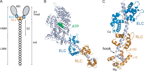
Structure of the myosin molecule. A, schematic diagram of the myosin molecule. The N-terminal region of the myosin heavy chain forms the globular motor domain (MD; gray), which contains the sites for ATP and actin binding. The ELC (blue) and the RLC (orange) stabilize a single α-helical polypeptide chain at the C terminus of the motor domain. The remainder of the heavy chain forms an ∼160-nm α-helical coiled-coil rod, which gives rise to the filamentous properties of class II myosins. Proteolysis of myosin produces a soluble subfragment (HMM), consisting of the S1 head and the adjacent S2 rod, and the insoluble light meromyosin (LMM) fragment responsible for myosin assembly. The molecular mass of the myosin heavy chain is ∼200 kDa; the RLC and ELC are each ∼20 kDa. B, ribbon representation of scallop myosin S1 (Protein Data Bank code 1QVI). The nucleotide (ATP; green) pocket is located at the lower end of a large cleft that serves as a communication pathway between actin and the nucleotide-binding site. C, ribbon diagram of the scallop regulatory domain (Protein Data Bank code 1WDC), with Ca2+ (cyan) bound to domain 1 of the ELC and Mg2+ (yellow) bound to the N terminus of the RLC. The α-helical myosin heavy chain (gray) has a sharp bend or hook near the C terminus (6, 7).
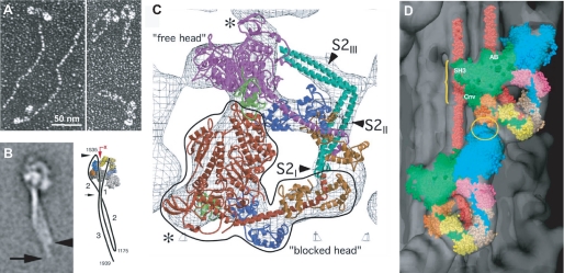
Inhibited configuration of the myosin molecule. A, electron micrographs of smooth muscle myosin rotary-shadowed with platinum at high (extended 6 S form) and low (folded 10 S form) ionic strength (13). B, electron microscopy of folded smooth muscle myosin molecules by negative staining and single-particle image processing (22). The accompanying diagram shows the two bends in the rod near residues 1175 and 1535 to form the 10 S structure. A photoactivated probe (the red arrow points to RLC Cys108) can cross-link the rod to the LC (43). C, electron cryomicroscopy of two-dimensional crystalline arrays of unphosphorylated smooth muscle HMM on a lipid monolayer surface (19). The outlined blocked head (red) interacts with the converter (green) and the ELC (blue) on the free head (magenta). The assignment for the S2 density is uncertain and more likely follows a path between the heads as indicated by the negatively stained molecules in B. D, atomic model of smooth muscle HMM (see C) fitted into the three-dimensional reconstruction of a tarantula muscle myosin filament obtained by electron cryomicroscopy and single-particle imaging techniques (25). The blocked and free heads are colored green and blue, respectively, in this representation. Regions of possible interactions are between the ELC and free heads (yellow ellipse) and between the blocked motor domain and S2 (yellow bracket). SH3, Src homology 3 domain; AB, actin binding; Cnv, converter.
References
-
- Rayment I., Rypniewski W. R., Schmidt-Bäse K., Smith R., Tomchick D. R., Benning M. M., Winkelmann D. A., Wesenberg G., Holden H. M. (1993) Science 261, 50–58 - PubMed
-
- Rayment I., Holden H. M., Whittaker M., Yohn C. B., Lorenz M., Holmes K. C., Milligan R. A. (1993) Science 261, 58–65 - PubMed
-
- Adelstein R. S., Conti M. A. (1975) Nature 256, 597–598 - PubMed
-
- Adelstein R. S., Eisenberg E. (1980) Annu. Rev. Biochem. 49, 921–956 - PubMed
Publication types
MeSH terms
Substances
Grants and funding
LinkOut - more resources
Full Text Sources
