. 2010 Apr 16;328(5976):372-6.
doi: 10.1126/science.1186068. Epub 2010 Mar 25.

Protein kinase C-theta mediates negative feedback on regulatory T cell function

Affiliations

Protein kinase C-theta mediates negative feedback on regulatory T cell function

Alexandra Zanin-Zhorov et al. Science. .

Erratum in

  • Science. 2010 May 21;328(5981):974. doi: 10.1126/science.328.5981.974-b

Abstract

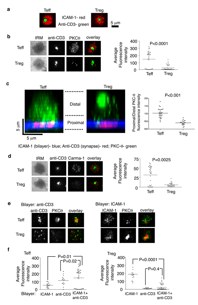
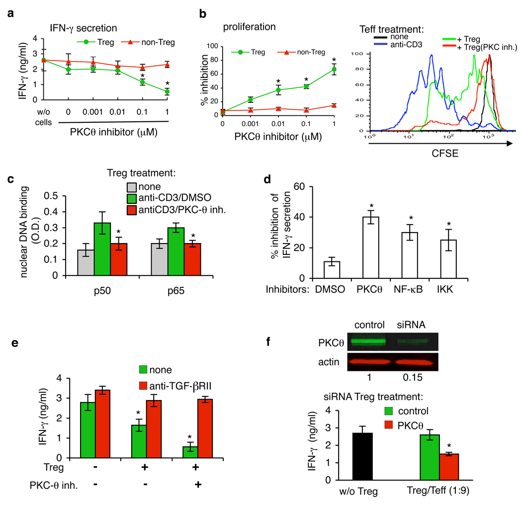
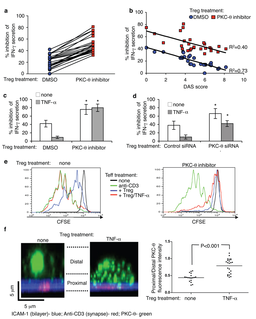
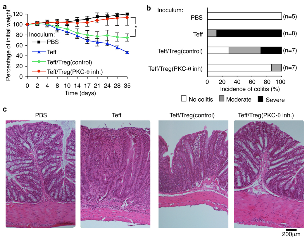
T cell receptor (TCR)-dependent regulatory T cell (Treg) activity controls effector T cell (Teff) function and is inhibited by the inflammatory cytokine tumor necrosis factor-alpha (TNF-alpha). Protein kinase C-theta (PKC-theta) recruitment to the immunological synapse is required for full Teff activation. In contrast, PKC-theta was sequestered away from the Treg immunological synapse. Furthermore, PKC-theta blockade enhanced Treg function, demonstrating PKC-theta inhibits Treg-mediated suppression. Inhibition of PKC-theta protected Treg from inactivation by TNF-alpha, restored activity of defective Treg from rheumatoid arthritis patients, and enhanced protection of mice from inflammatory colitis. Treg freed of PKC-theta-mediated inhibition can function in the presence of inflammatory cytokines and thus have therapeutic potential in control of inflammatory diseases.


Figures

Fig. 1
Fig. 1
Human Treg form altered IS. Human Teff and Treg were introduced into bilayers containing anti-CD3 (5 µg/ml) and ICAM-1 at 250 molecules/mm2 (A–D), fixed at 6 min and permeabilized, stained and imaged by TIRFM (A, B, D, E) or by confocal microscopy (C). (A) Localization of ICAM-1 (red) and anti-CD3 (green) in the IS. (B and D) Staining and average fluorescence intensity of PKC-θ (B) and Carma-1 (D). (C). Distribution of endogenous PKC-θ in cells. Histogram shows average of (bottom planes “proximal”/ upper planes “distal”) ratio of anti-PKCθ intensity per cell. The panels show representative images. (E and F) PKC-θ staining (E) and average fluorescence intensity (F) in Teff or Treg on bilayers containing only anti-CD3 (left) or ICAM-1 (right). Data are representative of three (A–D) or five (E–F) different experiments. P values were calculated by Mann-Whitney test.
Fig. 2
Fig. 2
PKC-θ inhibition up-regulates the suppressive function of Treg in vitro. (A and B) Treg and Teff were treated with PKC-θ inhibitor C20 at 0.001–1 µM for 30 min and washed three times. Treated cells were then mixed with CD4+ CD25 T (Teff) cells at a 1:9 ratio and plated on immobilized anti-CD3 mAb. (A) The supernatants were analyzed for IFN-γ after 24 hours by ELISA. (B)Cell proliferation was determined after 96 hours by Alamar Blue (left panel) or Treg were treated with 1 µM C20 for 30 min, washed, mixed with Teff and proliferation was assayed by CFSE (right panel) (C) Treg were treated with 1 µM of C20 for 30 min, washed, stimulated by anti-CD3 and p65/p50 specific binding to NF-κB consensus sequence was determined by ELISA. (D) Tregs were treated with 1 µM of NF-κB and IKK inhibitors, mixed with Teff and IFN-γ secretion was analyzed as in (A) after 48 hours. (E) Treg were treated with 1 µM of C20, mixed with Teff and cultured on anti-CD3 mAb with or without of neutralizing anti-TGF-β RII antibodies (20 µg/ml). IFN-γ secretion was analyzed as in (A) after 48 hours. (F) Treg were transfected with small interfering RNA (siRNA) targeting PKC-θ, or with control siRNA and plated on anti-CD3. After 48 hours PKC-θ expression was measured by Western blot analysis (top panel). siRNA-transfected Treg were mixed with CD4+ CD25 T (Teff) cells at 1:9 ratio and plated on immobilized anti-CD3 mAb. The supernatants were analyzed for IFN-γ after 48 hours. Average of four different experiments are shown for all panels. P values were calculated by t-test.
Fig. 3
Fig. 3
Inhibition of PKC-θ rescues and protects Treg function. (A and B) Freshly purified Treg from healthy donors and RA patients were treated or not with PKC-θ inhibitor C20 for 30 min at 1µM, washed three times, mixed with CD4+CD25 T cells at ratio 1:3, and plated on immobilized anti-CD3. The supernatants were analyzed for IFN-γ after 24–48 hours by ELISA. (C–E) Treg from healthy donors were treated with PKC-θ inhibitor C20 (C, E) or with PKC-θ siRNA (D) as described above, and co-cultured with CD4+ CD25 T cells with or without TNF-α (50 ng/ml). IFN-γ secretion was determined after 48 hours by ELISA (C and D) and proliferation was determined after 96 hours (E). (F) Untreated or TNF-α-treated (50 ng/ml, for 24 hr) Treg were introduced to bilayers with anti-CD3 and ICAM-1, fixed, and imaged by confocal microscopy. The panels show representative images with ICAM-1 (blue), anti-CD3 (red) and PKC-θ (green). Plots show the distribution of endogenous PKC-θ in cells. Each bar shows average of (bottom planes “synapse proximal”/ upper planes “synapse distal”) ratio of anti-PKCθ intensity per cell. Data points from three independent experiments are included in the analysis.
Fig. 4
Fig. 4
Treatment with PKC-θ inhibitor up-regulates Treg function in vivo. Colitis was induced in C57BL/10.PL TCRα−/− β−/− mice (31). Disease progression was monitored by (A) body weight loss, (B) colitis score and (C) histology. Numbers in parentheses indicate number of mice. Combined data of three independent experiments are presented. P values were calculated by t-test.

Comment in

  • Regulatory T-cell function in autoimmunity.
    Garcia-Valladares I, Espinoza LR. Garcia-Valladares I, et al. Immunotherapy. 2010 Jul;2(4):441-2. doi: 10.2217/imt.10.37. Immunotherapy. 2010. PMID: 20635997 No abstract available.

References

    1. Sakaguchi S, Yamaguchi T, Nomura T, Ono M. Cell. 2008 May 30;133:775. - PubMed
    1. Shevach EM, et al. Immunol Rev. 2006 Aug;212:60. - PubMed
    1. Zheng Y, Rudensky AY. Nat Immunol. 2007 May;8:457. - PubMed
    1. Flores-Borja F, Mauri C, Ehrenstein MR. Eur J Immunol. 2008 Apr;38:934. - PubMed
    1. Valencia X, et al. Blood. 2006 Jul 1;108:253. - PMC - PubMed

Publication types

MeSH terms