. 2010 Mar 26;327(5973):1638-42.
doi: 10.1126/science.1184429.

Shaping development of autophagy inhibitors with the structure of the lipid kinase Vps34

Affiliations

Shaping development of autophagy inhibitors with the structure of the lipid kinase Vps34

Simon Miller et al. Science. .

Abstract

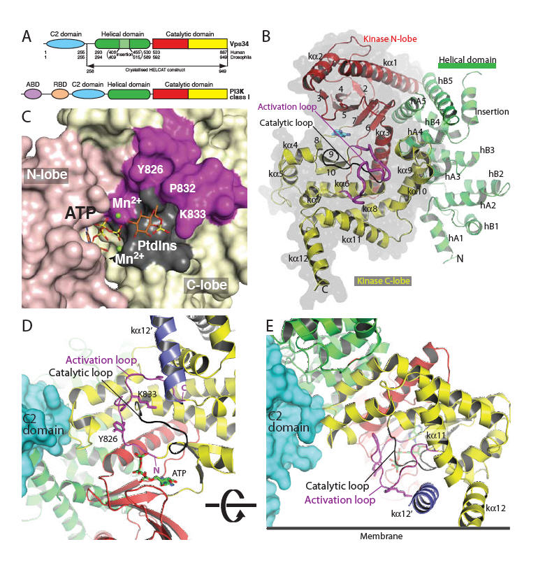
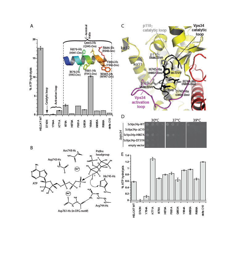
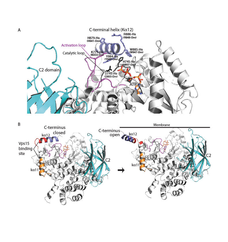
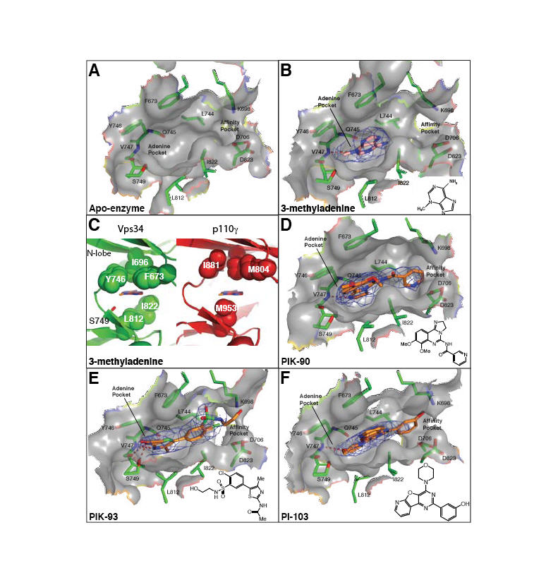
Phosphoinositide 3-kinases (PI3Ks) are lipid kinases with diverse roles in health and disease. The primordial PI3K, Vps34, is present in all eukaryotes and has essential roles in autophagy, membrane trafficking, and cell signaling. We solved the crystal structure of Vps34 at 2.9 angstrom resolution, which revealed a constricted adenine-binding pocket, suggesting the reason that specific inhibitors of this class of PI3K have proven elusive. Both the phosphoinositide-binding loop and the carboxyl-terminal helix of Vps34 mediate catalysis on membranes and suppress futile adenosine triphosphatase cycles. Vps34 appears to alternate between a closed cytosolic form and an open form on the membrane. Structures of Vps34 complexes with a series of inhibitors reveal the reason that an autophagy inhibitor preferentially inhibits Vps34 and underpin the development of new potent and specific Vps34 inhibitors.


Figures

Fig. 1
Fig. 1
Structure of Vps34 catalytic core (HELCAT). (A) Domain organisation of Vps34 and class I PI3Ks. (B) Overall fold of the DmVps34 HELCAT. (C) A view of the hook-shaped activation loop (magenta) encircling the catalytic loop (black). The C2 domain (cyan) is that of p110γ after superimposing DmVps34 residues 291-949 onto p110γ. The kα12′ helix (slate) is the C-terminal helix from the adjacent molecule in the crystal dimer. (D) The 2mFo-DFc electron density, contoured at 1.1σ, for the activation loop. (E) A model for PtdIns headgroup binding to Vps34, suggesting that Lys833-Dm (K771-Hs) interacts with the 1-phosphate. (F) The putative orientation of Vps34 on a membrane.
Fig. 2
Fig. 2
Essential structural elements for Vps34 catalysis. (A) The catalytic loop, the activation loop and the amphipathic C-terminal helix are critical for catalysis on PtdIns:PS vesicles. (B) Proposed catalytic mechanism of Vps34. (C) A close-up view of the proposed movements of catalytic loop residues His745-Hs and Asp743-Hs between the inactive and active conformations represented by the p110γ (grey) and DmVps34 crystal structure (black), respectively. (D) The ability of a yeast Vps34p-expressing plasmid to complement the growth defect of a Δvps34 yeast strain at elevated temperatures is impaired by deletion of the C-terminal helix (Vps34p ΔC10), a point mutation in this helix (ScH867A) or a mutation in the catalytic loop (ScD731N). (E) Basal ATPase activities in the absence of vesicles.
Fig. 3
Fig. 3
A model for Vps34 activation on membranes. (A) A close-up for the closed form of the enzyme in the cytosol. The C-terminal helix protects the phosphotransferase center from water. (B) The transition of the enzyme from a closed form in the cytosol (C-terminal helix in) to an open form with the C-terminal helix interacting with the membrane. The C2 domain (cyan) modelled as in Fig. 1. Vps15-interacting regions are classified as strong (red) or weak (orange) (27).
Fig. 4
Fig. 4
Inhibitor binding in the ATP pocket. (A-B,D-F) 2mFo-DFc electron densities are shown (contoured at 1.0σ). (C) A comparison of the ATP-binding pocket of the Vps34/3-MA complex (left, green) with p110γ (right, red). A ring of hydrophobic residues encircles 3-MA, and may provide specificity for Vps34. The p110γ structure is of the ATP/enzyme complex (PDB ID 1E8X), but a 3-MA has been modelled in the pocket as a reference point for comparison with the Vps34/3-MA complex. (G) Substitution of a cyclopentanecarboxamide in PT210 for the acetamide moiety of PIK93 reverses the specificity of the compound to selectively inhibit Vps34.

Comment in

References

    1. Engelman JA, Luo J, Cantley LC. Nat Rev Genet. 2006;7:606. - PubMed
    1. Schu PV, et al. Science. 1993;260:88. - PubMed
    1. Burda P, Padilla SM, Sarkar S, Emr SD. J Cell Sci. 2002;115:3889. - PubMed
    1. Vieira OV, et al. J Cell Biol. 2001;155:19. - PMC - PubMed
    1. Vergne I, et al. Proc Natl Acad Sci U S A. 2005;102:4033. - PMC - PubMed

Publication types

MeSH terms