. 2010 Jun;30(6):1189-95.
doi: 10.1161/ATVBAHA.110.205377. Epub 2010 Mar 25.

C-reactive protein-mediated vascular injury requires complement

Affiliations

C-reactive protein-mediated vascular injury requires complement

Fadi G Hage et al. Arterioscler Thromb Vasc Biol. 2010 Jun.

Abstract

Background: We previously demonstrated that vascular injury-induced neointima formation is exaggerated in human C-reactive protein (CRP) transgenic (CRPtg) compared to nontransgenic (NTG) mice. We now test the hypothesis that complement is required for this effect.

Methods and results: CRPtg and NTG with a normal complement system versus their counterparts lacking expression of complement component 3 (C3) protein (CRPtg/C3(-/-) and NTG/C3(-/-)) underwent carotid artery ligation. Twenty-eight days later, the injured vessels in CRPtg had thicker neointimas and more immunoreactive C3 in the surrounding adventitia compared with NTG. In CRPtg/C3(-/-), there was no increase in neointimal thickness compared with NTG or NTG/C3(-/-). Decreasing human CRP blood levels through administration of a selective antisense oligonucleotide eliminated the depletion of serum C3 associated with vascular injury and reduced immunoreactive C3 in the resultant lesions. In injured vessels, C3 colocalized with F4/80 (macrophage marker), and in vitro, human CRP elicited increased expression of C3 by bone marrow-derived macrophages.

Conclusions: Human CRP exaggeration of neointima formation in injured mouse carotid arteries associates with decreased circulating C3 and increased tissue-localized C3. C3 elimination or pharmacological reduction of human CRP prevents CRP-driven exacerbation of the injury response. In the CRPtg model system, mouse C3 is essential for the effect of human CRP.


Figures
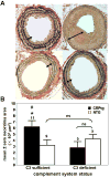
Figure 1
Figure 1
A. Representative light micrographs of Verhoeff's elastin stained sections of carotid arteries (taken 350 μm from the point of ligation) harvested 28 days after ligation injury from NTG versus CRPtg mice that were either C3 sufficient or C3 deficient. Note that the neointima (white double-headed arrows) of CRPtg is thicker than that of NTG if C3 is sufficient (upper right versus upper left panel, respectively), but this is not the case if C3 is deficient (lower right versus lower left panel, respectively). B. Cross-sectional areas of the neointimas of carotid arteries harvested 28 days after ligation injury from NTG versus CRPtg mice that were either C3 sufficient or C3 deficient. Note that the neointimal area (the average of sections taken 200, 350, and 500 μm from the point of ligation) of CRPtg is greater than NTG only if C3 is present. The numbers above the bars indicate the number of individual mouse arteries analyzed. *p<0.05 vs. NTG/C3 sufficient mice; # p<0.05 vs. CRPtg/C3 deficient mice; ns, not significant.
Figure 2
Figure 2
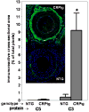
Immunoreactive complement C3 (green staining) and immunoreactive complement C5 (red staining) in sections of carotid arteries (taken 350 μm from the point of ligation) harvested 14 days after ligation injury. Immunoreactive C3, seen primarily in the adventitia, was found at higher levels in injured arteries of CRPtg than NTG mice (*, p<0.05 versus NTG). Immunoreactive C5 was present at lower levels, was localized in the neointima and did not differ between genotypes. The blue color indicates DAPI-stained cell nuclei. The data shown are means ± sems for sections taken from n=5 to 11 separate mice.
Figure 3
Figure 3
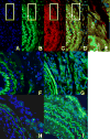
A-E. CRPtg carotid arteries harvested 14 days after injury were stained with DAPI (blue color) to reveal nuclei (panel A) and with fluorochrome labeled antibodies to reveal mouse C3 (green, panel B) and the F4/80 mouse macrophage marker (red, panel C). In the merged image (panel D) C3+/F4/80+ double-stained cells are seen to populate the adventitia. The area enclosed by the white rectangle is reproduced at higher magnification in panel E. F-H. Carotid arteries harvested 14 days after injury were stained with DAPI (blue color) to reveal nuclei and with fluorochrome labeled antibody to reveal human CRP (green). In both CRPtg (panel G) and CRPtg/C3-/- (panel I) abundant human CRP is seen, whereas none is seen in their NTG counterparts (panels F and G, respectively). Note that the elastic laminae are autofluorescent in the red and green channels.
Figure 4
Figure 4
The effect of human CRP on C3 and C5 mRNA expression by bone-marrow derived macrophages. In CRPtg carotid arteries harvested 24 h after injury (A), human CRP (green) colocalizes (large arrowhead) with mouse F4/80 (red, small arrows), indicating that CRP is associated with macrophages in vivo soon after vascular ligation. In NTG injured arteries (B), no human CRP is seen on macrophages surrounding the artery (EL = elastic laminae, Lu= vessel lumen, blue staining corresponds to DAPI-stained cell nuclei). Bone-marrow derived macrophages from CRPtg and NTG mice expressed increased levels of C3 (C) but not C5 (D) mRNA in response to 50μg/ml CRP (right panel). *p<0.05 vs. vehicle treated cells of the same genotype. The data shown are means ± sems for C3 and C5 mRNA levels for n=3 to 5 experiments.
Figure 5
Figure 5
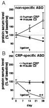
Lowering of human CRP blood levels abrogates depletion of mouse C3 after carotid artery ligation. A. Human CRP blood levels (open circles) rise ∼90% and mouse blood C3 (closed circles) falls ∼40% in CRPtg mice treated with a control scrambled antisense oligonucleotide. B. In CRPtg mice treated with the human CRP specific antisense oligonucleotide, human CRP level drops by more than 90% whereas there is no concomitant change in mouse C3. *p<0.05 vs. C3 levels in CRP-specific ASO treated mice, **p<0.05 vs. human CRP levels in non-specific ASO treated mice.
Figure 6
Figure 6
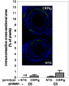
Immunoreactive complement in ligated carotid arteries (sections taken 350 μm from the point of ligation) at 28 days after injury in CRPtg and NTG mice pretreated for 2 weeks with a CRP specific antisense oligonucleotide that lowered serum CRP prior to carotid artery ligation. Immunoreactive C3 (green) and C5 (red) staining in the CRPtg were comparable to the NTG. Blue staining corresponds to cell nuclei (DAPI). The data shown are for sections taken from n=5 to 11 separate mice.

References

    1. Xing D, Hage FG, Chen YF, McCrory MA, Feng W, Skibinski GA, Majid-Hassan E, Oparil S, Szalai AJ. Exaggerated neointima formation in human C-reactive protein transgenic mice is IgG Fc receptor type I (Fc gamma RI)-dependent. The American journal of pathology. 2008;172:22–30. - PMC - PubMed
    1. Wang D, Oparil S, Chen YF, McCrory MA, Skibinski GA, Feng W, Szalai AJ. Estrogen treatment abrogates neointima formation in human C-reactive protein transgenic mice. Arteriosclerosis, thrombosis, and vascular biology. 2005;25:2094–2099. - PubMed
    1. Stein MP, Mold C, Du Clos TW. C-reactive protein binding to murine leukocytes requires Fc gamma receptors. J Immunol. 2000;164:1514–1520. - PubMed
    1. Lu J, Marnell LL, Marjon KD, Mold C, Du Clos TW, Sun PD. Structural recognition and functional activation of FcgammaR by innate pentraxins. Nature. 2008;456:989–992. - PMC - PubMed
    1. Hage FG, McCrory MA, Szalai AJ. C-reactive protein and cardiovascular disease: Lessons learned from studying genetically engineered mice. In: Nagasawa S, editor. C-Reactive Protein – New Research. Hauppauge NY: Nova Publishers; 2008. pp. 83–116.

Publication types

MeSH terms

Substances