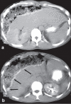
Spontaneous rupture of a giant hepatic hemangioma - sequential management with transcatheter arterial embolization and resection
- PMID: 20339183
- PMCID: PMC3016500
- DOI: 10.4103/1319-3767.61240
Spontaneous rupture of a giant hepatic hemangioma - sequential management with transcatheter arterial embolization and resection
Abstract
Hemangioma is the most common benign tumor of liver and is often asymptomatic. Spontaneous rupture is rare but has a catastrophic outcome if not promptly managed. Emergent hepatic resection has been the treatment of choice but has high operative mortality. Preoperative transcatheter arterial embolization (TAE) can significantly improve outcome in such patients. We report a case of spontaneous rupture of giant hepatic hemangioma that presented with abdominal pain and shock due to hemoperitoneum. Patient was successfully managed by TAE, followed by tumor resection. TAE is an effective procedure in symptomatic hemangiomas, and should be considered in such high risk patients prior to surgery.
Conflict of interest statement
Figures


References
-
- Brouwers MA, Peeters PM, de Jong KP, Haagsma EB, Klompmaker IJ, Bijleveld CM, et al. Surgical treatment of giant hemangioma of the liver. Br J Surg. 1997;84:314–6. - PubMed
-
- Cappellani A, Zanghi A, Di Vita M, Zanghi G, Tomarchio G, Petrillo G. Spontaneous rupture of a giant hemangioma of the liver. Ann Ital Chir. 2000;71:379–83. - PubMed
-
- Corigliano N, Mercantini P, Amodio PM, Balducci G, Caterino S, Ramacciato G, et al. Hemoperitoneum from a spontaneous rupture of a giant hemangioma of liver: Report of a case. Surg Today. 2003;33:459–63. - PubMed
-
- Ishak KG, Rabin L. Benign tumors of the liver. Med Clin North Am. 1975;59:995–1013. - PubMed
Publication types
MeSH terms
LinkOut - more resources
Full Text Sources
Medical
