

Titin diversity--alternative splicing gone wild
- PMID: 20339475
- PMCID: PMC2843904
- DOI: 10.1155/2010/753675
Titin diversity--alternative splicing gone wild
Abstract
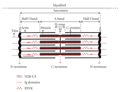
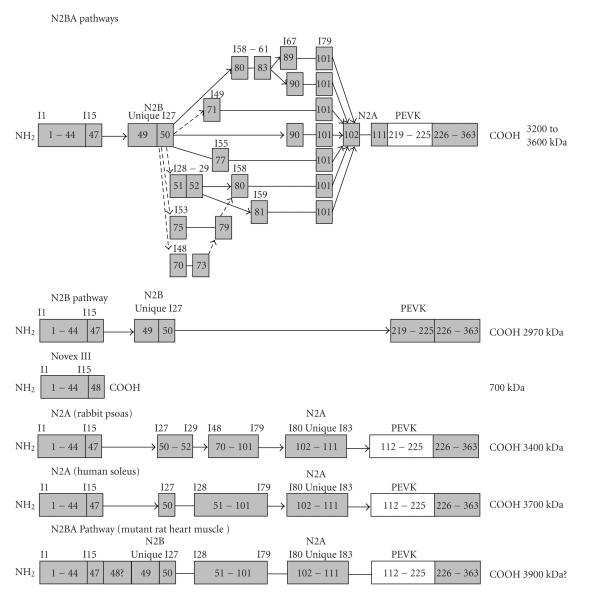
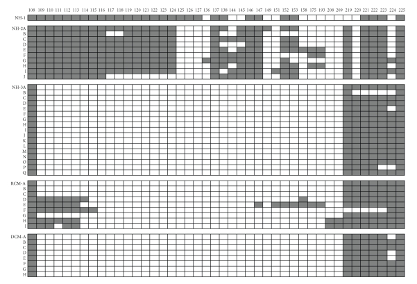
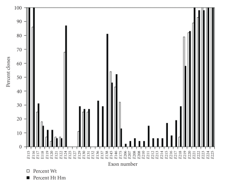
Titin is an extremely large protein found in highest concentrations in heart and skeletal muscle. The single mammalian gene is expressed in multiple isoforms as a result of alternative splicing. Although titin isoform expression is controlled developmentally and in a tissue specific manner, the vast number of potential splicing pathways far exceeds those described in any other alternatively spliced gene. Over 1 million human splice pathways for a single individual can be potentially derived from the PEVK region alone. A new splicing pattern for the human cardiac N2BA isoform type has been found in which the PEVK region includes only the N2B type exons. The alterations in splicing and titin isoform expression in human heart disease provide impetus for future detailed study of the splicing mechanisms for this giant protein.
Figures
References
-
- Labeit S, Kolmerer B, Linke WA. The giant protein titin: emerging roles in physiology and pathophysiology. Circulation Research. 1997;80(2):290–294. - PubMed
-
- Maruyama K. Connectin, an elastic protein from myofibrils. Journal of Biochemistry. 1976;80(2):405–407. - PubMed
-
- Furst DO, Osborn M, Nave R, Weber K. The organization of titin filaments in the half-sarcomere revealed by monoclonal antibodies in immunoelectron microscopy: a map of ten nonrepetitive epitopes starting at the Z-line extends close to the M line. Journal of Cell Biology. 1988;106(5):1563–1572. - PMC - PubMed
-
- Labeit S, Kolmerer B. Titins: giant proteins in charge of muscle ultrastructure and elasticity. Science. 1995;270(5234):293–296. - PubMed
Publication types
MeSH terms
Substances
Grants and funding
LinkOut - more resources
Full Text Sources
