. 2010 Mar 18:4:7.
doi: 10.3389/fnsys.2010.00007. eCollection 2010.

3D-Reconstructions and Virtual 4D-Visualization to Study Metamorphic Brain Development in the Sphinx Moth Manduca Sexta

Affiliations

3D-Reconstructions and Virtual 4D-Visualization to Study Metamorphic Brain Development in the Sphinx Moth Manduca Sexta

Wolf Huetteroth et al. Front Syst Neurosci. .

Abstract

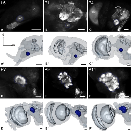
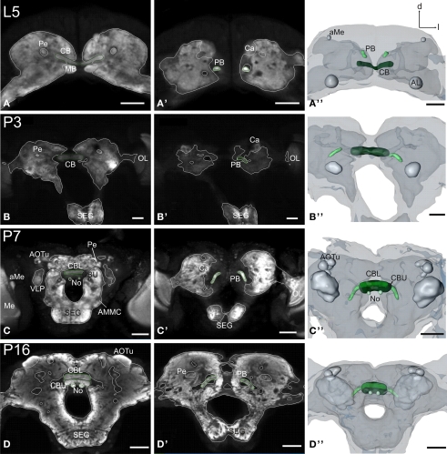
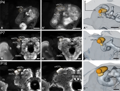
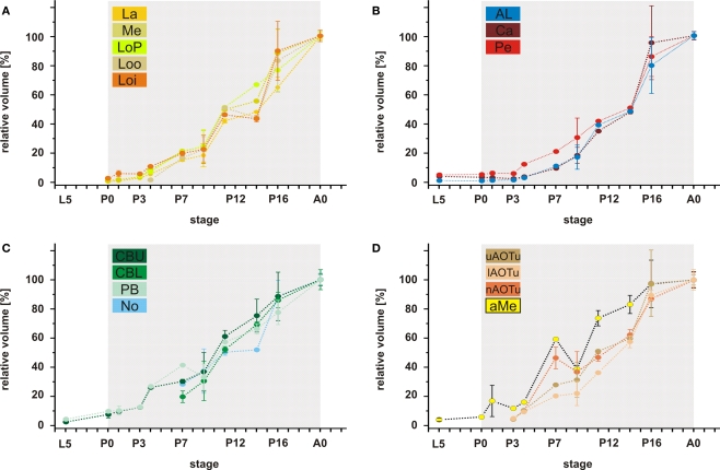
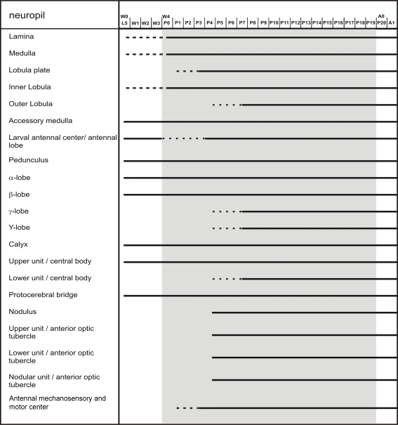
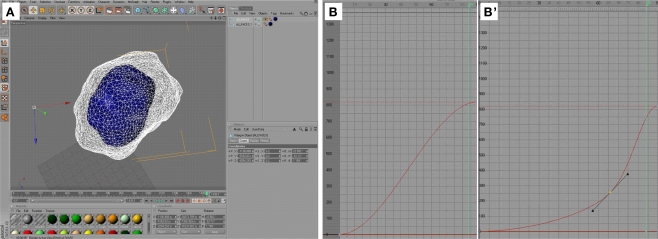
DURING METAMORPHOSIS, THE TRANSITION FROM THE LARVA TO THE ADULT, THE INSECT BRAIN UNDERGOES CONSIDERABLE REMODELING: new neurons are integrated while larval neurons are remodeled or eliminated. One well acknowledged model to study metamorphic brain development is the sphinx moth Manduca sexta. To further understand mechanisms involved in the metamorphic transition of the brain we generated a 3D standard brain based on selected brain areas of adult females and 3D reconstructed the same areas during defined stages of pupal development. Selected brain areas include for example mushroom bodies, central complex, antennal- and optic lobes. With this approach we eventually want to quantify developmental changes in neuropilar architecture, but also quantify changes in the neuronal complement and monitor the development of selected neuronal populations. Furthermore, we used a modeling software (Cinema 4D) to create a virtual 4D brain, morphing through its developmental stages. Thus the didactical advantages of 3D visualization are expanded to better comprehend complex processes of neuropil formation and remodeling during development. To obtain datasets of the M. sexta brain areas, we stained whole brains with an antiserum against the synaptic vesicle protein synapsin. Such labeled brains were then scanned with a confocal laser scanning microscope and selected neuropils were reconstructed with the 3D software AMIRA 4.1.

Keywords: Manduca; animation; brain; development; digital neuroanatomy; insect; neuropil.


Figures

Figure 1
Figure 1
Development of the adult optic lobe. Single optical sections in the frontal plane at different depths, anti-synapsin immunostaining (left two columns). Right column, 3D reconstructions (frontal views). (A–A′′) Last larval stage (L5): the larval optic center (LOC) is hardly distinguishable from the midbrain neuropil (gray line). The accessory medulla (aMe) is part of the LOC. IOA, inner optic anlage; OOA, outer optic anlage. (B–B′′) Stage P0: the ribbon-like adult neuropils lamina (La), medulla (Me) and lobula (Lo) show faint anti-synapsin staining. The aMe is separated from the midbrain neuropil and stays close to the antero-medial rim of the Me. Note the strong anti-synapsin immunoreactivity in the ventrolateral protocerebrum (VLP). (C–C′′) Stage P3: the La and Me further expand and the dorsal and ventral tips of the La bend behind the Me. The lobula plate (LoP) appears between Me and Lo. The anti-synapsin staining gains intensity in all neuropils; layers of the Me become recognizable. (D–D′′) Stage P7: the inner and outer lobula (Loi, Loo) can be separated. The La starts to exhibit its bowl-like appearance, the opening still tilted towards the anterior side. (E–E′′) Stage P14: all optic lobe neuropils reached their final adult positions. Orientation bars: d, dorsal; l, lateral; scale bars: 100 μm (A–B′′); 200 μm (C–E′′).
Figure 2
Figure 2
Development of the antennal lobe. Single optical sections in the frontal plane, anti-synapsin immunostaining (first and third row). Second and fourth row, 3D reconstructions (frontal views). (A–A) The larval antennal center (LAC) exhibits no glomerular organization in Manduca sexta L5 larva. (B–B) Strong anti-synapsin immunoreactivity is visible in the ventro-lateral protocerebrum (VLP), compared to the weak labeling in the LAC/AL. (C–C) Stage P4: the developing AL becomes recognizable in the anti-synapsin staining. Note the ventral position of the AL compared to stage P7. (D–D) Stage P7: glomerular structures surrounding a coarse neuropil become first visible. (E–E) Stage P9: single glomeruli become discernible structures and achieve their final position within the AL. The dorsal migration of the AL ends. (F–F) Stage P14: glomerular and AL volume increased compared to stage P9. Orientation bars: d, dorsal; m, medial; all scale bars: 100 μm.
Figure 3
Figure 3
Development of the mushroom body. Single optical sections in the frontal plane at different depths, anti-synapsin immunostaining (left two columns). Right column, 3D reconstructions (frontal views). (A–A′′) Rather thin α-and β-lobes (αL, βL) and a slender peduncle are characteristic for the larval (L5) mushroom body (MB). The peduncle of the larval MB is oriented antero-ventrally, with a slight inclination of the calyces (Ca) to the dorsal midline. While the αL points antero-dorsally, the βL extends medially in the horizontal plane. (B–B′′) early pupae (P1): the peduncle shows a straightened orientation. (C–C′′) Stage P4: the MB tilts from a ventro-dorsal orientation to a more antero-posterior orientation. (D–D′′) Stage P7: the MB is oriented antero-posteriorly and has massively increased in size compared to stage P4. Additionally to the αL and βL, the γ-lobe (γL) and the Y-lobe (YL) appear as protrusions on the MB. (E–E′′) Stage P16: the MB reached its final adult orientation and shape. Orientation bars: d, dorsal; m, medial; all scale bars: 100 μm.
Figure 4
Figure 4
Development of the central complex. Single optical sections in the frontal plane at different depths, anti-synapsin immunostaining (left two columns). Right column, 3D reconstructions (frontal views). (A–A′′) Central body (CB) and protocerebral bridge (PB) are present in the L5 larva. The PB consists of two separate neuropils and retains this layout up to adulthood. (B–B′′) Stage P3: the two parts of the PB elongate horizontally while the CB becomes thicker. (C–C′′) Stage P7: a lower (CBL) and upper (CBU) subunit are discernable. The noduli (No) appear antero-ventrally of the CB. The PB reaches its final position with its lateral ends pointing postero-ventrally. (D–D′′) Stage P16: the central complex has further increased in size. Orientation bars: d, dorsal; l, lateral; scale bars: 100 μm (A–B′′); 200 μm (C–D′′).
Figure 5
Figure 5
Development of the anterior optic tubercle. Single optical sections in the frontal plane at different depths, anti-synapsin immunostaining (left two columns). Right column, 3D reconstructions (frontal views). (A–A′′) Stage P4: all three subcompartments of the anterior optic tubercle (AOTu) become discernible structures in the antero-dorso-lateral midbrain: the upper (uAOTu), lower (lAOTu), and the nodular subunit (nAOTu). (B–B′′) Stage P7: independent of the rotation events of mushroom bodies (MBs) and the central complex, the subunits of the AOTu stay in their position and increased in size. (C–C′′) Stage 16: all subunits of the AOTu increased further in size. The uAOTu remains the largest subunit throughout metamorphic development. Orientation bars: d, dorsal; m, medial; all scale bars: 100 μm.
Figure 6
Figure 6
3D brain reconstructions of representative developmental stages. Most conspicuous are the prominent overall size increases (especially in the optic lobes) and the fusion of the subesophageal and supraesophageal ganglion. Note the outward tilting movement of the mushroom bodies, together with a backward rotation. These brains represent the reference points for the 4D visualization (Movie S1 in Supplementary Material). Anterior view; scale bar: 500 μm.
Figure 7
Figure 7
Relative volumes of the examined neuropils. (A) The optic lobe neuropils; (B) The antennal lobes and the mushroom bodies; (C) The central complex; (D) The anterior optic tubercle and the accessory medulla. For each data point values of two brains are used (only for P4; n = 1). For bilateral neuropils, the means of the volumes of the individual animal were used. Deviation bars, standard errors. La, lamina; Me, medulla; LoP, lobula plate; Loo, outer lobula; Loi, inner lobula; AL, antennal lobe; Ca, calyx; Pe, pedunculus; CBU, upper unit of the central body (CB); CBL, lower unit of the CB; PB, protocerebral bridge; No, nodule; uAOTu, upper anterior optic tubercle (AOTu); lAOTu, lower AOTu; nAOTu, nodular subunit of the AOTu; a Me, accessory medulla.
Figure 8
Figure 8
Scheme of brain neuropil development. The scheme, based on anti-synapsin immunostaining, reflects the developmental time course of the individual neuropils. Dotted lines represent the gradual appearance of a neuropil, or, in case of the larval antennal center, its transition into the antennal lobe.
Figure 9
Figure 9
Cinema 4D user interface – animation of the visualization. (A) Interactive mode in Cinema 4D; the center of the adult antennal lobe (A0, white, polygon grid view) is registered semi-automatically with the center of the P16 antennal lobe (blue, polygon surface view). Subsequently, the polygons are adjusted manually to match the shape of the corresponding preceding neuropil. (B–B) Detail of the timeline window; the f-curve (red) represents the relative position (y-axis) of a certain neuropil in relation to frame number (x-axis) between two key points. By default, this curve is sigmoidal (B) and needs to be adjusted manually by adding another key point (yellow, (B)).

Similar articles

Cited by

References

    1. Amos T. M., Gelman D. B., Mesce K. A. (1996). Steroid hormone fluctuations regulate ganglionic fusion during metamorphosis of the moth Manduca sexta. J. Insect Physiol. 42, 579–59110.1016/0022-1910(95)00127-1 - DOI
    1. Amos T. M., Mesce K. A. (1994). Reorganization of the ventral nerve cord in the moth Manduca sexta (L.) (Lepidoptera:Sphingidae). Int. J. Insect Morphol. Embryol. 23, 21–3710.1016/0020-7322(94)90013-2 - DOI
    1. Bell R. A., Joachim F. A. (1978). Techniques for rearing laboratory colonies of the tobacco hornworm, Manduca sexta, and pink ballworms. Ann. Entomol. Soc. Am. 69, 365–373
    1. Brandt R., Rohlfing T., Rybak J., Krofczik S., Maye A., Westerhoff M., Hege H. C., Menzel R. (2005). Three-dimensional average-shape atlas of the honeybee brain and its applications. J. Comp. Neurol. 492, 1–1910.1002/cne.20644 - DOI - PubMed
    1. Bullock T. H., Horridge G. A. (1965). Structure and Function in the Nervous Systems of Invertebrates, Vol. 2 San Francisco, Freeman

LinkOut - more resources