. 2010 Mar 3:4:3.
doi: 10.3389/neuro.06.003.2010. eCollection 2010.

3D Standard Brain of the Red Flour Beetle Tribolium Castaneum: A Tool to Study Metamorphic Development and Adult Plasticity

Affiliations

3D Standard Brain of the Red Flour Beetle Tribolium Castaneum: A Tool to Study Metamorphic Development and Adult Plasticity

David Dreyer et al. Front Syst Neurosci. .

Abstract

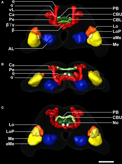
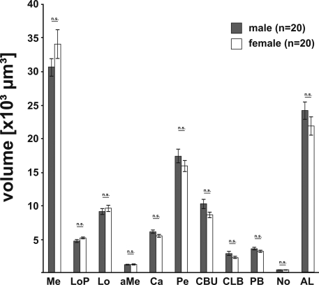
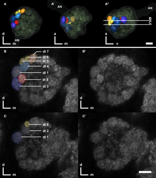
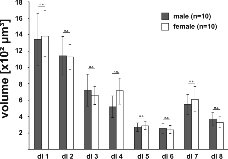
The red flour beetle Tribolium castaneum is emerging as a further standard insect model beside Drosophila. Its genome is fully sequenced and it is susceptible for genetic manipulations including RNA-interference. We use this beetle to study adult brain development and plasticity primarily with respect to the olfactory system. In the current study, we provide 3D standard brain atlases of freshly eclosed adult female and male beetles (A0). The atlases include eight paired and three unpaired neuropils including antennal lobes (ALs), optic lobe neuropils, mushroom body calyces and pedunculi, and central complex. For each of the two standard brains, we averaged brain areas of 20 individual brains. Additionally, we characterized eight selected olfactory glomeruli from 10 A0 female and male beetles respectively, which we could unequivocally recognize from individual to individual owing to their size and typical position in the ALs. In summary, comparison of the averaged neuropil volumes revealed no sexual dimorphism in any of the reconstructed neuropils in A0 Tribolium brains. Both, the female and male 3D standard brain are also used for interspecies comparisons, and, importantly, will serve as future volumetric references after genetical manipulation especially regarding metamorphic development and adult plasticity.

Keywords: antennal lobe; brain; coleoptera; digital neuroanatomy; insect; neuropil; olfactory system.


Figures

Figure 1
Figure 1
Confocal images of the individual T. castaneum brain stained with an antibody against synapsin which was used as template for the VIB protocol. (A, B) Single optical sections through the brain in the frontal (A) and horizontal plane (B). All manually labeled neuropil areas are shown as reconstructed in AMIRA. Labels of paired neuropils are only visualized in the left hemispheres, to provide a comparison to the unlabeled neuropils of the right hemispheres. (A) Frontal sections with the positions a–f from anterior to posterior and as described in (D). (B) Horizontal sections with the positions a´–c´ from dorsal to ventral as described in (D). (C) The color code of the labeled neuropils is consistent with Brandt et al. (2005), Kurylas et al. (2008), and el Jundi et al. (2009). AL, antennal lobe; Ca, Calyx; CBL, lower unit of the central body; CBU, upper unit of the central body; aMe, accessory medulla; Lo, lobula; LoP, lobula plate; Me, medulla; No, nodulus; PB, protocerebral bridge; and Pe, pedunculus with lobes. (D) Volume rendered (Voltex) view of the template brain from dorsal and frontal. The sections (a–f) and (a–c) represent the positions of the optical sections in (A,B). Orientation bars, p, posterior; m, median; d, dorsal. All scale bars, 50 μm.
Figure 2
Figure 2
3D reconstruction of the male template brain of T. castaneum in (A) anterior (B) ventral, and (C) posterior view. The neuropils were visualized with the AMIRA tools SurfaceGen and Surfaceview. Note that the α and α´ lobes of the medial lobes can be clearly distinguished. In the medial lobes, the β-lobes are visible, while the second protrusion represents the β´- and the γ-lobes which were not discernable in the reconstruction. vL, vertical lobe. See Figure 1 for color code and abbreviations. Scale bar, 50 μm.
Figure 3
Figure 3
3D male standard brain of T. castaneum calculated from 20 individual brains by using the VIB protocol. (A–C) Surface reconstructions of all 18 averaged labels in (A) anterior (B) ventral, and (C) posterior view. The brain surface (as in A´–C´) is not labeled. See Figure 1 for color code. (A–C) 3D visualization of the corresponding average intensity map by direct volume rendering with (A) anterior (B) ventral, and (C) posterior view, using non-rigid transformation. (D, E) Direct volume rendered view of the resulting average label images from (D) anterior and (E) posterior, exhibiting deviations primarily in the lobes of the pedunculi of the MBs. Scale bar, 50 μm.
Figure 4
Figure 4
Comparison of neuropil volumes between male (gray) and female (white) brains. The data revealed no significant difference (two-tailed Student's t-test) between the volumes of the corresponding neuropils of females and males. Bars, standard error; n.s., not significant.
Figure 5
Figure 5
Right antennal lobe of a male T. castaneum brain. (A–A′)Anterior (A), ventral (A), and lateral view (A′′) of 3D-reconstructed glomeruli including the antennal nerve (AN, transparent). The eight color coded dorso-lateral glomeruli (dl-1 to 8, compare with B,C) can be unequivocally identified in 75% of all preparations. Other glomeruli are depicted in transparent green; the displayed outline of the AL is shown in transparent gray. The transparent encasement around the AL represents the shape of the whole AL. The vertical bars in (A′′) display section levels of (B,C) and (B,C′), respectively. Orientation bars: a, anterior; d, dorsal; m, medial; v, ventral. (B,C) Frontal confocal sections through the antennal lobe according to (A′′). The eight dorso-lateral glomeruli are manually labeled as reconstructed in AMIRA. (B,C) Confocal sections through the antennal lobe corresponding to (B,C). All scale bars: 10 μm.
Figure 6
Figure 6
Comparison of the volumes of the eight identified glomeruli between right male (gray; n = 10) and right female (white; n = 10) ALs, revealed no significant difference between the sexes (two-tailed student t-test). Bars, standard error; n.s., not significant.

References

    1. Anton S., Homberg U. (1999). Antennal lobe structure. In Insect Olfaction, Hansson B. S., ed. (Berlin, Springer; ), pp. 97–124
    1. Arnaud L., Lognay G., Verscheure M., Leenaers L., Gaspar C., Haubruge E. (2002). Is dimethyldecanal a common aggregation pheromone of Tribolium flour beetles? J. Chem. Ecol. 28, 523–53210.1023/A:1014587927784 - DOI - PubMed
    1. Arnold G., Masson C., Budharugsa S. (1984). Demonstration of a sexual dimorphism in the olfactory pathways of the drones of Apis mellifica L. (Hymenoptera, Apidae). Experientia 40, 723–72510.1007/BF01949744 - DOI
    1. Barth M., Heisenberg M. (1997). Vision affects mushroom bodies and central complex in Drosophila melanogaster. Learn. Mem. 4, 219–22910.1101/lm.4.2.219 - DOI - PubMed
    1. Barth M., Hirsch H. V., Meinertzhagen I. A., Heisenberg M. (1997). Experience-dependent developmental plasticity in the optic lobe of Drosophila melanogaster. J. Neurosci. 17, 1493–1504 - PMC - PubMed