. 2010 Mar 22;5(3):e9776.
doi: 10.1371/journal.pone.0009776.

The lectin ArtinM induces recruitment of rat mast cells from the bone marrow to the peritoneal cavity

Affiliations

The lectin ArtinM induces recruitment of rat mast cells from the bone marrow to the peritoneal cavity

Patricia Andressa de Almeida Buranello et al. PLoS One. .

Abstract

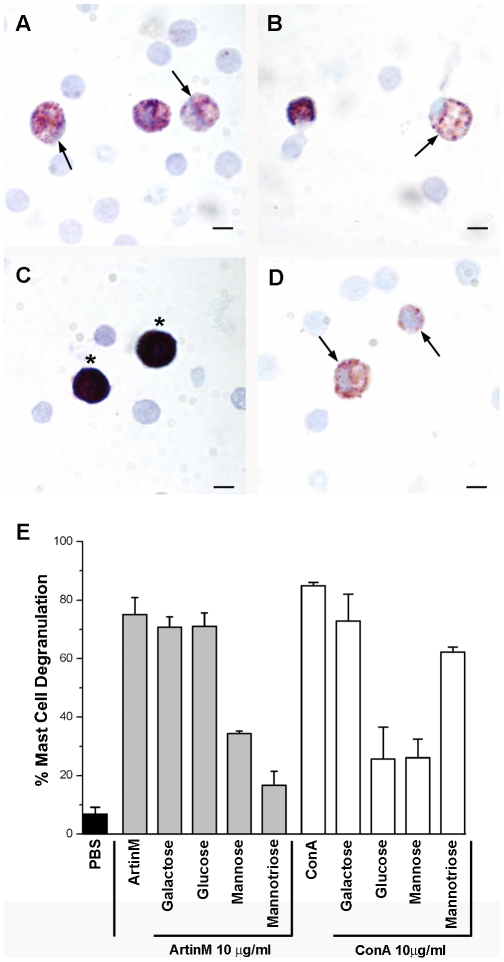
Background: The D-mannose binding lectin ArtinM is known to recruit neutrophils, to degranulate mast cells and may have potential therapeutic applications. However, the effect of ArtinM on mast cell recruitment has not been investigated.

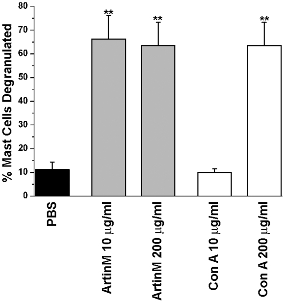
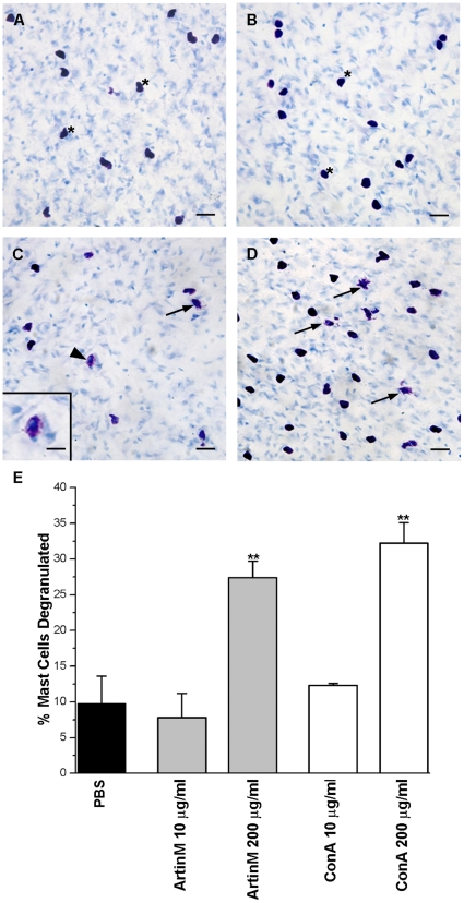
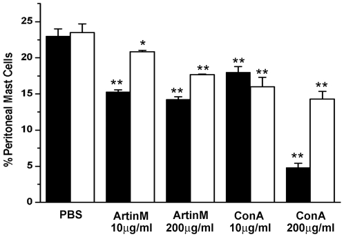
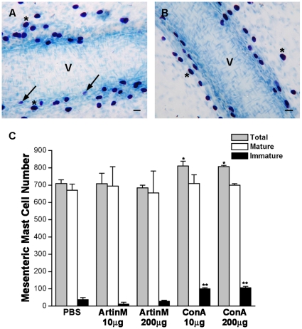
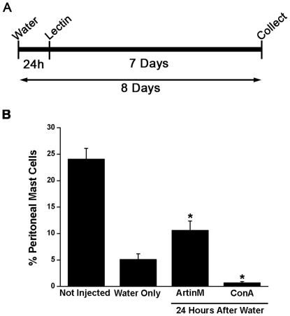
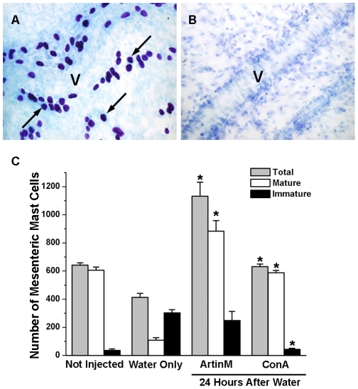
Methodology: Male Wistar rats were injected i.p. with ArtinM or ConA (control). The ability of the lectin to degranulate peritoneal and mesenteric mast cells was examined. Recruitment of mast cells to the peritoneal cavity and mesentery after ArtinM injection was examined with or without depletion of peritoneal mast cells by distilled water.

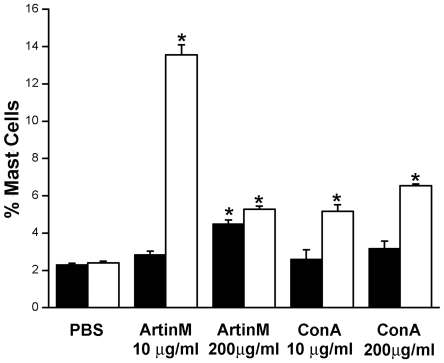
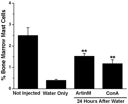
Results: ArtinM degranulated both peritoneal and mesentery mast cells in vitro. Three days after i.p. injection of the lectin there were reduced numbers of mast cells in the peritoneal lavage, while at 7 days post injection of ArtinM, the number of peritoneal mast cells was close to control values. Since immature mast cells are recruited from the bone marrow, the effect of the lectin on bone marrow mast cells was examined. Injection of ArtinM resulted in an increased number of mast cells in the bone marrow. To determine if degranulation of mast cells in the peritoneal cavity was required for the increase in bone marrow mast cells, the peritoneal cavity was depleted of mast cells with ultrapure water. Exposure to ArtinM increased the number of mast cells in the bone marrow of rats depleted of peritoneal mast cells.

Conclusions: The ArtinM induced recruitment of mast cells from the bone marrow to the peritoneal cavity may partially explain the therapeutic actions of ArtinM.


Conflict of interest statement

Competing Interests: The authors have declared that no competing interests exist.

Figures

Figure 1
Figure 1. ArtinM and ConA degranulate peritoneal mast cells in vitro in a lectin specific manner.
The total cell population from the peritoneal lavage was incubated with 10 µg of ArtinM (A) or ConA (B). Control cells were incubated in PBS (C). Compound 48/80 (D) was used as a positive control for degranulation. Stained with Toluidine blue. (Arrows, degranulated mast cells; Asterisks, intact mast cells) Bars = 10 µm. Quantification of peritoneal mast cell degranulation (E). The data shown is the average±SD of 3 separate experiments.
Figure 2
Figure 2. ArtinM and Con A also degranulate mast cells in vivo.
The peritoneal lavage was examined 1 hr after i.p. injection of 10 µg of either lectin. There was no significant difference in the ability the two lectins to degranulate mast cells. **p≤0.01. The data shown is the average±SD of 3 separate experiments.
Figure 3
Figure 3. ArtinM and ConA also degranulate mesenteric mast cells in vitro only at high concentrations.
Mesenteries were incubated with 10 µg/ml ArtinM (A), 10 µg/ml ConA (B), 200 µg/ml ArtinM (C) or 200 µg/ml ConA (D). (Arrows, degranulated mast cells; *, non degranulated mast cells). Bar = 20 µm. Inset (C): Details of degranulated mast cell indicated by arrowhead. Bar = 10 µm Stain: Toluidine blue. Quantification of mesenteric mast cell degranulation by ArtinM or ConA (E). At 200 µg of either ArtinM or ConA there was significant (**p≤0.01) degranulation of mesenteric mast cells. The data shown is the average±SD of 3 separate experiments.
Figure 4
Figure 4. Con A-FITC binds preferentially to the extracellular matrix.
Rats were injected i.p. with 10 µg Con A-FITC (A. Fluorescence image, B. DIC image) or with 200 µg Con A-FITC. (C. Fluorescence image, D. DIC image). (Mast cells, arrows).
Figure 5
Figure 5. Intraperitoneal injection of ArtinM and ConA reduces the percentage of metachromatic mast cells in the peritoneal lavage.
Animals were injected i.p. with 4 ml of PBS containing either 10 µg or 200 µg of ArtinM or ConA, and the percent of metachromatic mast cells present in the peritoneal lavage was examined at 3 and 7 days post injection. The results shown are the average±SD of 3 separate experiments. 3 days post injection: ▪; 7 days post injection: □. *p≤0.05, **p≤0.01.
Figure 6
Figure 6. ConA, but not ArtinM recruits immature mast cells to the mesentery.
In the mesentery, 7 days after i.p. injection of 200 µg of Con A immature mast cells (arrows) with few metachromatic granules as well as mature mast cells (*) were found next to a blood vessel (A). In animals injected with PBS, only mature mast cells (*) were seen along blood vessels (B). Stain: Toluidine blue. (C) Seven days after i.p. injection of 10 µg or 200 µg of ArtinM or ConA the total number of mesenteric mast cells as well as the number of mature and immature mast cells was determined. *p≤0.05. The results shown are the average±SD of 3 separate experiments.
Figure 7
Figure 7. Intraperitoneal injection of lectin increases the percent of mast cells in the bone marrow.
Animals were injected i.p. with Artin M or ConA. At 3 (▪) or 7 (□) days after injection mast cells were immunomagnetically isolated from the bone marrow using mAb AA4 and the percent of mast cells determined. *p≤0.05. The results shown are the average±SD of 3 separate experiments.
Figure 8
Figure 8. ArtinM recruits mast cells to the peritoneal cavity in mast cell depleted animals.
Animals were injected i.p. with purified water to deplete the peritoneal cavity of mast cells and 24 h later received an i.p. injection of 4 ml of PBS containing 10 µg of ArtinM or ConA. The peritoneal lavage was collected 7 days after lectin injection (A). The percent of peritoneal mast cells was determined under the various conditions (B). *p≤0.05. The results shown are the average±SD of 3 separate experiments.
Figure 9
Figure 9. Intraperitoneal lectin injection recruits mast cells to the mesentery in mast cell depleted animals.
Animals were injected i.p. with purified water to deplete the peritoneal cavity of mast cells and 24 h later received an i.p. injection containing 10 µg of ArtinM or ConA. The mesentery was collected 7 days after lectin injection. In uninjected animals (A) the mesentery was replete with mature mast cells. Many lying adjacent to blood vessels (V). Twenty four hours after injection of purified water (B) no mast cells could be seen in the mesentery. The total number of mesenteric mast cells as well as the number of mature and immature mesenteric mast cells was determined under the various conditions (C). *p≤0.05. The results shown are the average±SD of 3 separate experiments.
Figure 10
Figure 10. Intraperitoneal Injection of either ArtinM or ConA resulted in a significant increase in the percent of mast cells in the bone marrow.
Eight days after injection of water and 7 days after injection of 10 µg of ArtinM or ConA, mast cells were immunomagnetically isolated from the bone marrow with mAb AA4 conjugated magnetic beads. **p≤0,01. The results shown are the average±SD of 3 separate experiments.
Figure 11
Figure 11. ArtinM increases the number of bone marrow derived mast cells recruited to the peritoneal cavity.
The peritoneal cavity was depleted of mast cells by injection of ultrapure water prior to the infusion of immunomagnetically isolated bone marrow derived mast cells labeled with CellTracker Red CMTPX™ and i.p. injection of ArtinM. At 24 and 48 h after infusion, the presence of fluorescently labeled mast cells in the peritoneal lavage was analysed by FACS. Each data point represents the average number of cells±S.D. from 3 separate experiments. 100,000 cells were counted per time point in each experiment. (▪ with ArtinM; □ without ArtinM).

References

    1. Panunto-Castelo A, Souza MA, Roque-Barreira MC, Silva JS. KM+, a lectin from Artocarpus integrifolia, induces IL-12 p40 production by macrophages and switches from type 2 to type 1 cell-mediated immunity against Leishmania major antigens, resulting in BALB/c mice resistance to infection. Glycobiology. 2001;11:1035–1042. - PubMed
    1. Toledo KA, Scwartz C, Oliveira AF, Conrado MCAV, Bernardes ES, et al. Neutrophil activation induced by ArtinM: Release of inflammatory mediators and enhancement of effector functions. Immunology Letters. 2009;123:14–20. - PubMed
    1. Moreno AN, Jamur MC, Oliver C, Roque-Barreira MC. Mast cell degranulation induced by lectins: effect on neutrophil recruitment. Int Arch Allergy Immunol. 2003;132:221–230. - PubMed
    1. Teixeira CR, Cavassani KA, Gomes RB, Teixeira MJ, Roque-Barreira MC, et al. Potential of KM+ lectin in immunization against Leishmania amazonensis infection. Vaccine. 2006;24:3001–3008. - PubMed
    1. Coltri KC, Oliveira LL, Pinzan CF, Vendruscolo PE, Martinez R, et al. Therapeutic administration of KM+ lectin protects mice against Paracoccidioides brasiliensis infection via interleukin-12 production in a Toll-Like receptor 2-dependent mechanism. Am J Pathol. 2008;173:423–432. - PMC - PubMed

Publication types