Review
. 2010 Jul;67(13):2195-213.
doi: 10.1007/s00018-010-0324-8. Epub 2010 Mar 26.

The elegans of spindle assembly

Affiliations
Review

The elegans of spindle assembly

Thomas Müller-Reichert et al. Cell Mol Life Sci. 2010 Jul.

Abstract

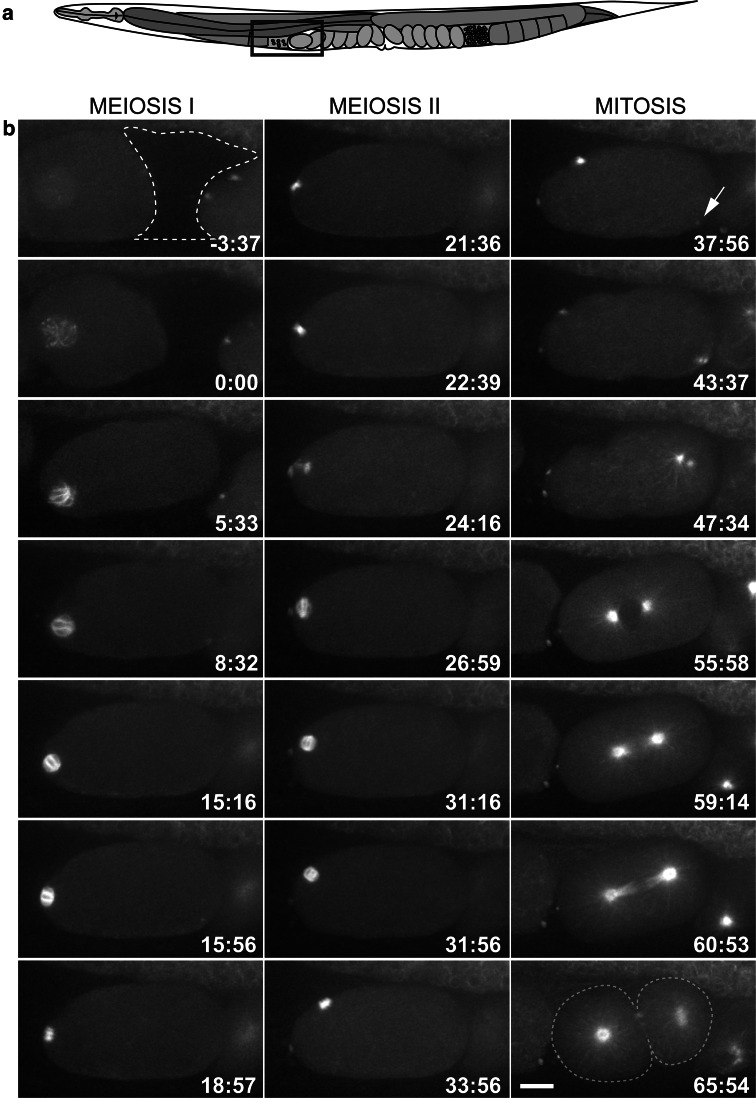
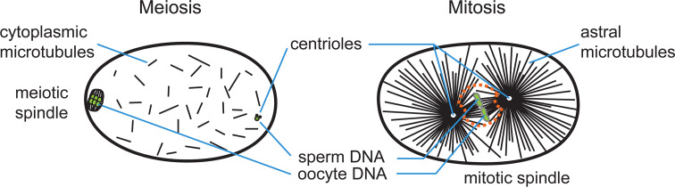
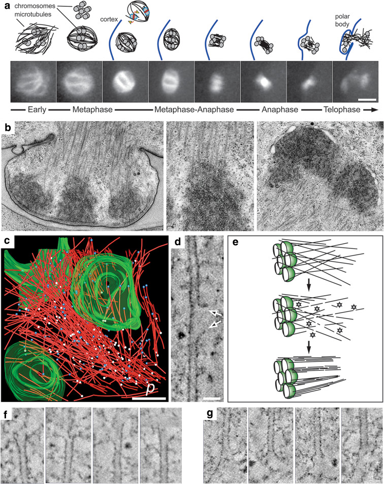
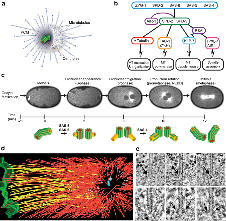
The Caenorhabditis elegans one-cell embryo is a powerful system in which to study microtubule organization because this large cell assembles both meiotic and mitotic spindles within the same cytoplasm over the course of 1 h in a stereotypical manner. The fertilized oocyte assembles two consecutive acentrosomal meiotic spindles that function to reduce the replicated maternal diploid set of chromosomes to a single-copy haploid set. The resulting maternal DNA then unites with the paternal DNA to form a zygotic diploid complement, around which a centrosome-based mitotic spindle forms. The early C. elegans embryo is amenable to live-cell imaging and electron tomography, permitting a detailed structural comparison of the meiotic and mitotic modes of spindle assembly.


Figures

Fig. 1
Fig. 1
Development of the one-cell C. elegans embryo as viewed by in utero-imaging of a worm expressing GFP::β-tubulin. a Schematic drawing of an adult hermaphrodite. The box depicts the region of the uterus imaged. b Microtubules are observed by fluorescent light microscopy around the oocyte nucleus (t = 0) before it enters the spermatheca (dashed line). Anterior is left in all panels. The oocyte usually enters the spermatheca within 5 min, at which time fertilization occurs and a pointed bipolar array becomes visible. Each round of meiosis takes approximately 20 min. At the end of meiosis II (t = 37:56), the centrosomal microtubules become visible (arrow), and centrosome separation begins soon after. The mitotic spindle forms in the center of the embryo and the spindle skews towards the posterior in anaphase to produce two daughter cells of unequal size. Scale bar is 10 μm in b
Fig. 2
Fig. 2
Schematic comparison of spindle assembly in meiosis and mitosis in the early C. elegans embryo. Chromatin is shown in green. The nuclear envelope (orange dashes) persists in the early stages of mitotic spindle assembly, but disappears by anaphase. Microtubules initially invade the nuclear space through polar fenestrae
Fig. 3
Fig. 3
Acentrosomal spindle organization in C. elegans female meiosis. a Schematic representation (above) of the first meiotic division (β-tubulin::GFP; lower panels). Microtubules form around the chromatin and become organized into a bipolar array (only three bivalents are drawn). At this stage, KLP-18 is required for microtubule bundling and pole focusing. Chromosomes align on the metaphase plate in a rosette pattern (shown above), and KLP-19 (red strip) is implicated in providing an anti-poleward force that could exert a torque on the paired chromosomes to ensure their proper alignment (yellow arrows). The redistribution of microtubules in metaphase-anaphase and telophase is represented schematically. By telophase, microtubules begin to accumulate around chromatin to assemble the second meiotic spindle. b Electron micrographs showing the formation of the first polar body with an enlarged view of a kinetochore region (mid panel). c Three-dimensional reconstruction of one half of a meiotic wild-type spindle (microtubules in red, pole–proximal ends as white spheres, pole–distal ends as blue spheres, chromatin in green; p is spindle pole, from [132]). d Tomographic slice showing lateral disruption of the lattice of a spindle microtubule (arrows). e Model explaining the role of katanin (shown as stars) in female meiotic spindle assembly. One half spindle is shown; chromatin is green. Katanin converts a few long microtubules into many short microtubule fragments that may become bundled and arranged into the bipolar array. f Gallery of microtubule pole–distal, and g gallery of microtubule pole–proximal ends showing a variety of open, flared morphologies. Scale bars are 5 μm in a, 500 nm in c, and 50 nm in d
Fig. 4
Fig. 4
Mitotic spindle organization in the C. elegans embryo. a Schematic representation of the C. elegans centrosome, with a pair of centrioles surrounded by pericentriolar material (PCM) and minus-end nucleated/anchored microtubules. b Functional levels of the centrosome. Cartoon illustrating core proteins responsible for centriole duplication (first level) and PCM assembly (second level). Proteins of the third level influence microtubule nucleation, polymerization and depolymerization. The aurora kinase AIR-1 is involved at multiple steps, in both the maturation of the centrosome and the regulation of downstream effectors. c Ultrastructural pathway of centriole duplication in C. elegans early embryos (modified from [68]). d Partial reconstruction of a metaphase half-spindle. Microtubules that contact the chromosomes (green) are designated kinetochore microtubules (shown in yellow). Other microtubules are displayed in red. Microtubule ends in the spindle pole, surrounding the centriole pair (blue cylinders), are indicated by white spheres. e Kinetochore microtubules have an open, flared plus-end morphology (upper row). About 80% of the microtubules in the centrosome (lower row) have closed, capped minus ends (arrowhead), although some microtubules with open ends (arrows) are observed (modified from [71])

Similar articles

Cited by

References

    1. Kimble JE, White JG. On the control of germ cell development in Caenorhabditis elegans . Dev Biol. 1981;81:208–219. doi: 10.1016/0012-1606(81)90284-0. - DOI - PubMed
    1. Miller MA, Nguyen VQ, Lee MH, Kosinski M, Schedl T, Caprioli RM, Greenstein D. A sperm cytoskeletal protein that signals oocyte meiotic maturation and ovulation. Science. 2001;291:2144–2147. doi: 10.1126/science.1057586. - DOI - PubMed
    1. Miller MA, Ruest PJ, Kosinski M, Hanks SK, Greenstein D. An Eph receptor sperm-sensing control mechanism for oocyte meiotic maturation in Caenorhabditis elegans . Genes Dev. 2003;17:187–200. doi: 10.1101/gad.1028303. - DOI - PMC - PubMed
    1. Ward S, Carrel JS. Fertilization and sperm competition in the nematode Caenorhabditis elegans . Dev Biol. 1979;73:304–321. doi: 10.1016/0012-1606(79)90069-1. - DOI - PubMed
    1. Yang HY, McNally K, McNally FJ. MEI-1/katanin is required for translocation of the meiosis I spindle to the oocyte cortex in C. elegans . Dev Biol. 2003;260:245–259. doi: 10.1016/S0012-1606(03)00216-1. - DOI - PubMed

Publication types

LinkOut - more resources